3iap: Difference between revisions
No edit summary |
No edit summary |
||
| Line 3: | Line 3: | ||
<StructureSection load='3iap' size='340' side='right'caption='[[3iap]], [[Resolution|resolution]] 2.00Å' scene=''> | <StructureSection load='3iap' size='340' side='right'caption='[[3iap]], [[Resolution|resolution]] 2.00Å' scene=''> | ||
== Structural highlights == | == Structural highlights == | ||
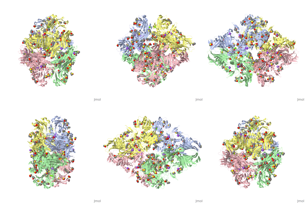
<table><tr><td colspan='2'>[[3iap]] is a 4 chain structure with sequence from [ | <table><tr><td colspan='2'>[[3iap]] is a 4 chain structure with sequence from [https://en.wikipedia.org/wiki/Ecoli Ecoli]. Full crystallographic information is available from [http://oca.weizmann.ac.il/oca-bin/ocashort?id=3IAP OCA]. For a <b>guided tour on the structure components</b> use [https://proteopedia.org/fgij/fg.htm?mol=3IAP FirstGlance]. <br> | ||
</td></tr><tr id='ligand'><td class="sblockLbl"><b>[[Ligand|Ligands:]]</b></td><td class="sblockDat"><scene name='pdbligand=BTB:2-[BIS-(2-HYDROXY-ETHYL)-AMINO]-2-HYDROXYMETHYL-PROPANE-1,3-DIOL'>BTB</scene>, <scene name='pdbligand=DMS:DIMETHYL+SULFOXIDE'>DMS</scene>, <scene name='pdbligand=MG:MAGNESIUM+ION'>MG</scene>, <scene name='pdbligand=NA:SODIUM+ION'>NA</scene></td></tr> | </td></tr><tr id='ligand'><td class="sblockLbl"><b>[[Ligand|Ligands:]]</b></td><td class="sblockDat" id="ligandDat"><scene name='pdbligand=BTB:2-[BIS-(2-HYDROXY-ETHYL)-AMINO]-2-HYDROXYMETHYL-PROPANE-1,3-DIOL'>BTB</scene>, <scene name='pdbligand=DMS:DIMETHYL+SULFOXIDE'>DMS</scene>, <scene name='pdbligand=MG:MAGNESIUM+ION'>MG</scene>, <scene name='pdbligand=NA:SODIUM+ION'>NA</scene></td></tr> | ||
<tr id='related'><td class="sblockLbl"><b>[[Related_structure|Related:]]</b></td><td class="sblockDat">[[1dp0|1dp0]], [[3dyp|3dyp]], [[3dym|3dym]], [[3iaq|3iaq]]</td></tr> | <tr id='related'><td class="sblockLbl"><b>[[Related_structure|Related:]]</b></td><td class="sblockDat"><div style='overflow: auto; max-height: 3em;'>[[1dp0|1dp0]], [[3dyp|3dyp]], [[3dym|3dym]], [[3iaq|3iaq]]</div></td></tr> | ||
<tr id='gene'><td class="sblockLbl"><b>[[Gene|Gene:]]</b></td><td class="sblockDat">lacZ ([ | <tr id='gene'><td class="sblockLbl"><b>[[Gene|Gene:]]</b></td><td class="sblockDat">lacZ ([https://www.ncbi.nlm.nih.gov/Taxonomy/Browser/wwwtax.cgi?mode=Info&srchmode=5&id=83333 ECOLI])</td></tr> | ||
<tr id='activity'><td class="sblockLbl"><b>Activity:</b></td><td class="sblockDat"><span class='plainlinks'>[ | <tr id='activity'><td class="sblockLbl"><b>Activity:</b></td><td class="sblockDat"><span class='plainlinks'>[https://en.wikipedia.org/wiki/Beta-galactosidase Beta-galactosidase], with EC number [https://www.brenda-enzymes.info/php/result_flat.php4?ecno=3.2.1.23 3.2.1.23] </span></td></tr> | ||
<tr id='resources'><td class="sblockLbl"><b>Resources:</b></td><td class="sblockDat"><span class='plainlinks'>[ | <tr id='resources'><td class="sblockLbl"><b>Resources:</b></td><td class="sblockDat"><span class='plainlinks'>[https://proteopedia.org/fgij/fg.htm?mol=3iap FirstGlance], [http://oca.weizmann.ac.il/oca-bin/ocaids?id=3iap OCA], [https://pdbe.org/3iap PDBe], [https://www.rcsb.org/pdb/explore.do?structureId=3iap RCSB], [https://www.ebi.ac.uk/pdbsum/3iap PDBsum], [https://prosat.h-its.org/prosat/prosatexe?pdbcode=3iap ProSAT]</span></td></tr> | ||
</table> | </table> | ||
== Evolutionary Conservation == | == Evolutionary Conservation == | ||
Revision as of 15:09, 13 October 2021
E. coli (lacZ) beta-galactosidase (E416Q)E. coli (lacZ) beta-galactosidase (E416Q)
Structural highlights
Evolutionary Conservation Check, as determined by ConSurfDB. You may read the explanation of the method and the full data available from ConSurf. Publication Abstract from PubMedVariants of beta-galactosidase with Valine and with Glutamine replacing Glutamate-416 did not have a Mg(2+) bound at the active site even at high Mg(2+) concentrations (200 mM). They had low catalytic activity and the pH profiles were very different from those of the native enzyme. In addition, substrates, substrate analogs, transition state analogs and galactose bound very poorly. However, the orientation and conformation of the Mg(2+) ligands (residues 416, 418, and 461) as well as the B-factors of these three side chains did not change significantly. The structures, conformations and B-factors of other active site residues were also essentially unchanged. These studies show that the active site Mg(2+) is not necessary for structure and is, therefore, mainly important for modulating the chemistry and mediating the interactions between the active site components. Studies of Glu-416 Variants of beta-Galactosidase (E. coli) Show that the Active Site Mg(2+) is Not Important for Structure and Indicate that the Main Role of Mg (2+) is to Mediate Optimization of Active Site Chemistry.,Lo S, Dugdale ML, Jeerh N, Ku T, Roth NJ, Huber RE Protein J. 2009 Nov 21. PMID:19936901[1] From MEDLINE®/PubMed®, a database of the U.S. National Library of Medicine. References
|
| ||||||||||||||||||||||
