1div: Difference between revisions
No edit summary |
No edit summary |
||
| Line 3: | Line 3: | ||
<StructureSection load='1div' size='340' side='right'caption='[[1div]], [[Resolution|resolution]] 2.60Å' scene=''> | <StructureSection load='1div' size='340' side='right'caption='[[1div]], [[Resolution|resolution]] 2.60Å' scene=''> | ||
== Structural highlights == | == Structural highlights == | ||
<table><tr><td colspan='2'>[[1div]] is a 1 chain structure with sequence from [ | <table><tr><td colspan='2'>[[1div]] is a 1 chain structure with sequence from [https://en.wikipedia.org/wiki/Geobacillus_stearothermophilus Geobacillus stearothermophilus]. Full crystallographic information is available from [http://oca.weizmann.ac.il/oca-bin/ocashort?id=1DIV OCA]. For a <b>guided tour on the structure components</b> use [https://proteopedia.org/fgij/fg.htm?mol=1DIV FirstGlance]. <br> | ||
</td></tr><tr id='resources'><td class="sblockLbl"><b>Resources:</b></td><td class="sblockDat"><span class='plainlinks'>[ | </td></tr><tr id='resources'><td class="sblockLbl"><b>Resources:</b></td><td class="sblockDat"><span class='plainlinks'>[https://proteopedia.org/fgij/fg.htm?mol=1div FirstGlance], [http://oca.weizmann.ac.il/oca-bin/ocaids?id=1div OCA], [https://pdbe.org/1div PDBe], [https://www.rcsb.org/pdb/explore.do?structureId=1div RCSB], [https://www.ebi.ac.uk/pdbsum/1div PDBsum], [https://prosat.h-its.org/prosat/prosatexe?pdbcode=1div ProSAT]</span></td></tr> | ||
</table> | </table> | ||
== Function == | == Function == | ||
[[ | [[https://www.uniprot.org/uniprot/RL9_GEOSE RL9_GEOSE]] Binds to the 23S rRNA. | ||
== Evolutionary Conservation == | == Evolutionary Conservation == | ||
[[Image:Consurf_key_small.gif|200px|right]] | [[Image:Consurf_key_small.gif|200px|right]] | ||
Revision as of 12:27, 21 July 2021
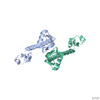
RIBOSOMAL PROTEIN L9RIBOSOMAL PROTEIN L9
Structural highlights
Function[RL9_GEOSE] Binds to the 23S rRNA. Evolutionary Conservation Check, as determined by ConSurfDB. You may read the explanation of the method and the full data available from ConSurf. Publication Abstract from PubMedThe crystal structure of protein L9 from the Bacillus stearothermophilus ribosome has been determined at 2.8 A resolution using X-ray diffraction methods. This primary RNA-binding protein has a highly elongated and unusual structure consisting of two separated domains joined by a long exposed alpha-helix. Conserved, positively charged and aromatic amino acids on the surfaces of both domains probably represent the sites of specific interactions with 23S rRNA. Comparisons with other prokaryotic L9 sequences show that while the length of the connecting alpha-helix is invariant, the sequence within the exposed central region is not conserved. This suggests that the alpha-helix has an architectural role and serves to fix the relative separation and orientation of the N- and C-terminal domains within the ribosome. The N-terminal domain has structural homology to the smaller ribosomal proteins L7/L12 and L30, and the eukaryotic RNA recognition motif (RRM). Crystal structure of prokaryotic ribosomal protein L9: a bi-lobed RNA-binding protein.,Hoffman DW, Davies C, Gerchman SE, Kycia JH, Porter SJ, White SW, Ramakrishnan V EMBO J. 1994 Jan 1;13(1):205-12. PMID:8306963[1] From MEDLINE®/PubMed®, a database of the U.S. National Library of Medicine. See AlsoReferences |
| ||||||||||||||