1hzh: Difference between revisions
No edit summary |
No edit summary |
||
| Line 3: | Line 3: | ||
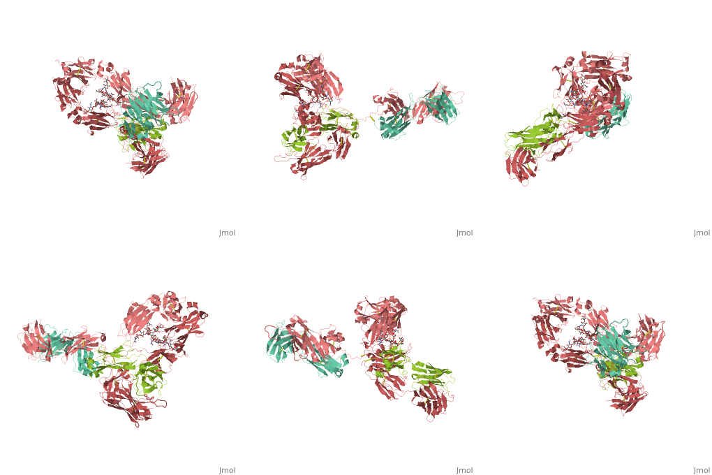
<StructureSection load='1hzh' size='340' side='right'caption='[[1hzh]], [[Resolution|resolution]] 2.70Å' scene=''> | <StructureSection load='1hzh' size='340' side='right'caption='[[1hzh]], [[Resolution|resolution]] 2.70Å' scene=''> | ||
== Structural highlights == | == Structural highlights == | ||
<table><tr><td colspan='2'>[[1hzh]] is a 4 chain structure. The September 2001 RCSB PDB [ | <table><tr><td colspan='2'>[[1hzh]] is a 4 chain structure with sequence from [https://en.wikipedia.org/wiki/Human Human]. The September 2001 RCSB PDB [https://pdb.rcsb.org/pdb/static.do?p=education_discussion/molecule_of_the_month/index.html Molecule of the Month] feature on ''Antibodies'' by David S. Goodsell is [https://dx.doi.org/10.2210/rcsb_pdb/mom_2001_9 10.2210/rcsb_pdb/mom_2001_9]. Full crystallographic information is available from [http://oca.weizmann.ac.il/oca-bin/ocashort?id=1HZH OCA]. For a <b>guided tour on the structure components</b> use [https://proteopedia.org/fgij/fg.htm?mol=1HZH FirstGlance]. <br> | ||
</td></tr><tr id='ligand'><td class="sblockLbl"><b>[[Ligand|Ligands:]]</b></td><td class="sblockDat"><scene name='pdbligand=BMA:BETA-D-MANNOSE'>BMA</scene>, <scene name='pdbligand=FUC:ALPHA-L-FUCOSE'>FUC</scene>, <scene name='pdbligand=GAL:BETA-D-GALACTOSE'>GAL</scene>, <scene name='pdbligand=MAN:ALPHA-D-MANNOSE'>MAN</scene>, <scene name='pdbligand=NAG:N-ACETYL-D-GLUCOSAMINE'>NAG</scene></td></tr> | </td></tr><tr id='ligand'><td class="sblockLbl"><b>[[Ligand|Ligands:]]</b></td><td class="sblockDat" id="ligandDat"><scene name='pdbligand=BMA:BETA-D-MANNOSE'>BMA</scene>, <scene name='pdbligand=FUC:ALPHA-L-FUCOSE'>FUC</scene>, <scene name='pdbligand=GAL:BETA-D-GALACTOSE'>GAL</scene>, <scene name='pdbligand=MAN:ALPHA-D-MANNOSE'>MAN</scene>, <scene name='pdbligand=NAG:N-ACETYL-D-GLUCOSAMINE'>NAG</scene></td></tr> | ||
<tr id='resources'><td class="sblockLbl"><b>Resources:</b></td><td class="sblockDat"><span class='plainlinks'>[ | <tr id='resources'><td class="sblockLbl"><b>Resources:</b></td><td class="sblockDat"><span class='plainlinks'>[https://proteopedia.org/fgij/fg.htm?mol=1hzh FirstGlance], [http://oca.weizmann.ac.il/oca-bin/ocaids?id=1hzh OCA], [https://pdbe.org/1hzh PDBe], [https://www.rcsb.org/pdb/explore.do?structureId=1hzh RCSB], [https://www.ebi.ac.uk/pdbsum/1hzh PDBsum], [https://prosat.h-its.org/prosat/prosatexe?pdbcode=1hzh ProSAT]</span></td></tr> | ||
</table> | </table> | ||
== Evolutionary Conservation == | == Evolutionary Conservation == | ||
| Line 30: | Line 30: | ||
*[[Antibody|Antibody]] | *[[Antibody|Antibody]] | ||
*[[Antibody 3D structures|Antibody 3D structures]] | *[[Antibody 3D structures|Antibody 3D structures]] | ||
*[[ | *[[3D structures of human antibody|3D structures of human antibody]] | ||
== References == | == References == | ||
<references/> | <references/> | ||
| Line 38: | Line 36: | ||
</StructureSection> | </StructureSection> | ||
[[Category: Antibodies]] | [[Category: Antibodies]] | ||
[[Category: Human]] | |||
[[Category: Large Structures]] | [[Category: Large Structures]] | ||
[[Category: RCSB PDB Molecule of the Month]] | [[Category: RCSB PDB Molecule of the Month]] | ||
Revision as of 12:51, 12 May 2021
CRYSTAL STRUCTURE OF THE INTACT HUMAN IGG B12 WITH BROAD AND POTENT ACTIVITY AGAINST PRIMARY HIV-1 ISOLATES: A TEMPLATE FOR HIV VACCINE DESIGNCRYSTAL STRUCTURE OF THE INTACT HUMAN IGG B12 WITH BROAD AND POTENT ACTIVITY AGAINST PRIMARY HIV-1 ISOLATES: A TEMPLATE FOR HIV VACCINE DESIGN
Structural highlights
Evolutionary Conservation Check, as determined by ConSurfDB. You may read the explanation of the method and the full data available from ConSurf. Publication Abstract from PubMedWe present the crystal structure at 2.7 angstrom resolution of the human antibody IgG1 b12. Antibody b12 recognizes the CD4-binding site of human immunodeficiency virus-1 (HIV-1) gp120 and is one of only two known antibodies against gp120 capable of broad and potent neutralization of primary HIV-1 isolates. A key feature of the antibody-combining site is the protruding, finger-like long CDR H3 that can penetrate the recessed CD4-binding site of gp120. A docking model of b12 and gp120 reveals severe structural constraints that explain the extraordinary challenge in eliciting effective neutralizing antibodies similar to b12. The structure, together with mutagenesis studies, provides a rationale for the extensive cross-reactivity of b12 and a valuable framework for the design of HIV-1 vaccines capable of eliciting b12-like activity. Crystal structure of a neutralizing human IGG against HIV-1: a template for vaccine design.,Saphire EO, Parren PW, Pantophlet R, Zwick MB, Morris GM, Rudd PM, Dwek RA, Stanfield RL, Burton DR, Wilson IA Science. 2001 Aug 10;293(5532):1155-9. PMID:11498595[1] From MEDLINE®/PubMed®, a database of the U.S. National Library of Medicine. See AlsoReferences
|
| ||||||||||||||||
