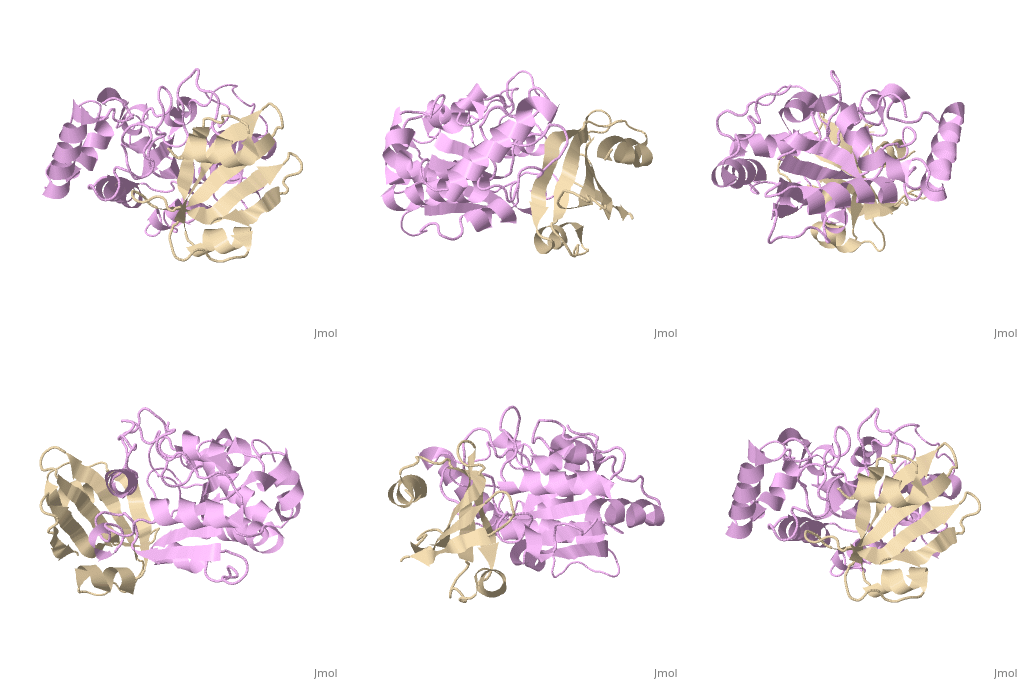
1ugh: Difference between revisions
No edit summary |
No edit summary |
||
| Line 1: | Line 1: | ||
==CRYSTAL STRUCTURE OF HUMAN URACIL-DNA GLYCOSYLASE IN COMPLEX WITH A PROTEIN INHIBITOR: PROTEIN MIMICRY OF DNA== | ==CRYSTAL STRUCTURE OF HUMAN URACIL-DNA GLYCOSYLASE IN COMPLEX WITH A PROTEIN INHIBITOR: PROTEIN MIMICRY OF DNA== | ||
<StructureSection load='1ugh' size='340' side='right' caption='[[1ugh]], [[Resolution|resolution]] 1.90Å' scene=''> | <StructureSection load='1ugh' size='340' side='right'caption='[[1ugh]], [[Resolution|resolution]] 1.90Å' scene=''> | ||
== Structural highlights == | == Structural highlights == | ||
<table><tr><td colspan='2'>[[1ugh]] is a 2 chain structure with sequence from [http://en.wikipedia.org/wiki/Bppb2 Bppb2] and [http://en.wikipedia.org/wiki/Human Human]. Full crystallographic information is available from [http://oca.weizmann.ac.il/oca-bin/ocashort?id=1UGH OCA]. For a <b>guided tour on the structure components</b> use [http://oca.weizmann.ac.il/oca-docs/fgij/fg.htm?mol=1UGH FirstGlance]. <br> | <table><tr><td colspan='2'>[[1ugh]] is a 2 chain structure with sequence from [http://en.wikipedia.org/wiki/Bppb2 Bppb2] and [http://en.wikipedia.org/wiki/Human Human]. Full crystallographic information is available from [http://oca.weizmann.ac.il/oca-bin/ocashort?id=1UGH OCA]. For a <b>guided tour on the structure components</b> use [http://oca.weizmann.ac.il/oca-docs/fgij/fg.htm?mol=1UGH FirstGlance]. <br> | ||
</td></tr><tr id='activity'><td class="sblockLbl"><b>Activity:</b></td><td class="sblockDat"><span class='plainlinks'>[http://en.wikipedia.org/wiki/Uridine_nucleosidase Uridine nucleosidase], with EC number [http://www.brenda-enzymes.info/php/result_flat.php4?ecno=3.2.2.3 3.2.2.3] </span></td></tr> | </td></tr><tr id='activity'><td class="sblockLbl"><b>Activity:</b></td><td class="sblockDat"><span class='plainlinks'>[http://en.wikipedia.org/wiki/Uridine_nucleosidase Uridine nucleosidase], with EC number [http://www.brenda-enzymes.info/php/result_flat.php4?ecno=3.2.2.3 3.2.2.3] </span></td></tr> | ||
<tr id='resources'><td class="sblockLbl"><b>Resources:</b></td><td class="sblockDat"><span class='plainlinks'>[http://oca.weizmann.ac.il/oca-docs/fgij/fg.htm?mol=1ugh FirstGlance], [http://oca.weizmann.ac.il/oca-bin/ocaids?id=1ugh OCA], [http://pdbe.org/1ugh PDBe], [http://www.rcsb.org/pdb/explore.do?structureId=1ugh RCSB], [http://www.ebi.ac.uk/pdbsum/1ugh PDBsum]</span></td></tr> | <tr id='resources'><td class="sblockLbl"><b>Resources:</b></td><td class="sblockDat"><span class='plainlinks'>[http://oca.weizmann.ac.il/oca-docs/fgij/fg.htm?mol=1ugh FirstGlance], [http://oca.weizmann.ac.il/oca-bin/ocaids?id=1ugh OCA], [http://pdbe.org/1ugh PDBe], [http://www.rcsb.org/pdb/explore.do?structureId=1ugh RCSB], [http://www.ebi.ac.uk/pdbsum/1ugh PDBsum], [http://prosat.h-its.org/prosat/prosatexe?pdbcode=1ugh ProSAT]</span></td></tr> | ||
</table> | </table> | ||
== Disease == | == Disease == | ||
| Line 14: | Line 15: | ||
Check<jmol> | Check<jmol> | ||
<jmolCheckbox> | <jmolCheckbox> | ||
<scriptWhenChecked>select protein; define ~consurf_to_do selected; consurf_initial_scene = true; script "/wiki/ConSurf/ug/1ugh_consurf.spt"</scriptWhenChecked> | <scriptWhenChecked>; select protein; define ~consurf_to_do selected; consurf_initial_scene = true; script "/wiki/ConSurf/ug/1ugh_consurf.spt"</scriptWhenChecked> | ||
<scriptWhenUnchecked>script /wiki/extensions/Proteopedia/spt/initialview01.spt</scriptWhenUnchecked> | <scriptWhenUnchecked>script /wiki/extensions/Proteopedia/spt/initialview01.spt</scriptWhenUnchecked> | ||
<text>to colour the structure by Evolutionary Conservation</text> | <text>to colour the structure by Evolutionary Conservation</text> | ||
| Line 31: | Line 32: | ||
==See Also== | ==See Also== | ||
*[[DNA glycosylase|DNA glycosylase]] | *[[DNA glycosylase 3D structures|DNA glycosylase 3D structures]] | ||
*[[Uracil | *[[Uracil glycosylase inhibitor|Uracil glycosylase inhibitor]] | ||
== References == | == References == | ||
<references/> | <references/> | ||
| Line 41: | Line 40: | ||
[[Category: Bppb2]] | [[Category: Bppb2]] | ||
[[Category: Human]] | [[Category: Human]] | ||
[[Category: Large Structures]] | |||
[[Category: Uridine nucleosidase]] | [[Category: Uridine nucleosidase]] | ||
[[Category: Arvai, A S]] | [[Category: Arvai, A S]] | ||
Revision as of 16:34, 17 July 2019
CRYSTAL STRUCTURE OF HUMAN URACIL-DNA GLYCOSYLASE IN COMPLEX WITH A PROTEIN INHIBITOR: PROTEIN MIMICRY OF DNACRYSTAL STRUCTURE OF HUMAN URACIL-DNA GLYCOSYLASE IN COMPLEX WITH A PROTEIN INHIBITOR: PROTEIN MIMICRY OF DNA
Structural highlights
Disease[UNG_HUMAN] Defects in UNG are a cause of immunodeficiency with hyper-IgM type 5 (HIGM5) [MIM:608106]. A rare immunodeficiency syndrome characterized by normal or elevated serum IgM levels with absence of IgG, IgA, and IgE. It results in a profound susceptibility to bacterial infections.[1] [2] Function[UNG_HUMAN] Excises uracil residues from the DNA which can arise as a result of misincorporation of dUMP residues by DNA polymerase or due to deamination of cytosine. [UNGI_BPPB2] This protein binds specifically and reversibly to the host uracil-DNA glycosylase, preventing removal of uracil residues from PBS2 DNA by the host uracil-excision repair system. Evolutionary Conservation Check, as determined by ConSurfDB. You may read the explanation of the method and the full data available from ConSurf. Publication Abstract from PubMedUracil-DNA glycosylase inhibitor (Ugi) is a B. subtilis bacteriophage protein that protects the uracil-containing phage DNA by irreversibly inhibiting the key DNA repair enzyme uracil-DNA glycosylase (UDG). The 1.9 A crystal structure of Ugi complexed to human UDG reveals that the Ugi structure, consisting of a twisted five-stranded antiparallel beta sheet and two alpha helices, binds by inserting a beta strand into the conserved DNA-binding groove of the enzyme without contacting the uracil specificity pocket. The resulting interface, which buries over 1200 A2 on Ugi and involves the entire beta sheet and an alpha helix, is polar and contains 22 water molecules. Ugi binds the sequence-conserved DNA-binding groove of UDG via shape and electrostatic complementarity, specific charged hydrogen bonds, and hydrophobic packing enveloping Leu-272 from a protruding UDG loop. The apparent mimicry by Ugi of DNA interactions with UDG provides both a structural mechanism for UDG binding to DNA, including the enzyme-assisted expulsion of uracil from the DNA helix, and a crystallographic basis for the design of inhibitors with scientific and therapeutic applications. Crystal structure of human uracil-DNA glycosylase in complex with a protein inhibitor: protein mimicry of DNA.,Mol CD, Arvai AS, Sanderson RJ, Slupphaug G, Kavli B, Krokan HE, Mosbaugh DW, Tainer JA Cell. 1995 Sep 8;82(5):701-8. PMID:7671300[3] From MEDLINE®/PubMed®, a database of the U.S. National Library of Medicine. See AlsoReferences
|
| ||||||||||||||||
