1hzm: Difference between revisions
No edit summary |
No edit summary |
||
| Line 1: | Line 1: | ||
==STRUCTURE OF ERK2 BINDING DOMAIN OF MAPK PHOSPHATASE MKP-3: STRUCTURAL INSIGHTS INTO MKP-3 ACTIVATION BY ERK2== | ==STRUCTURE OF ERK2 BINDING DOMAIN OF MAPK PHOSPHATASE MKP-3: STRUCTURAL INSIGHTS INTO MKP-3 ACTIVATION BY ERK2== | ||
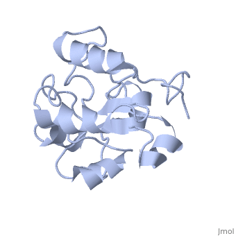
<StructureSection load='1hzm' size='340' side='right' caption='[[1hzm]], [[NMR_Ensembles_of_Models | 1 NMR models]]' scene=''> | <StructureSection load='1hzm' size='340' side='right' caption='[[1hzm]], [[NMR_Ensembles_of_Models | 1 NMR models]]' scene=''> | ||
== Structural highlights == | == Structural highlights == | ||
<table><tr><td colspan='2'>[[1hzm]] is a 1 chain structure with sequence from [http://en.wikipedia.org/wiki/Human Human]. Full experimental information is available from [http://oca.weizmann.ac.il/oca-bin/ocashort?id=1HZM OCA]. For a <b>guided tour on the structure components</b> use [http://oca.weizmann.ac.il/oca-docs/fgij/fg.htm?mol=1HZM FirstGlance]. <br> | <table><tr><td colspan='2'>[[1hzm]] is a 1 chain structure with sequence from [http://en.wikipedia.org/wiki/Human Human]. Full experimental information is available from [http://oca.weizmann.ac.il/oca-bin/ocashort?id=1HZM OCA]. For a <b>guided tour on the structure components</b> use [http://oca.weizmann.ac.il/oca-docs/fgij/fg.htm?mol=1HZM FirstGlance]. <br> | ||
</td></tr><tr id='resources'><td class="sblockLbl"><b>Resources:</b></td><td class="sblockDat"><span class='plainlinks'>[http://oca.weizmann.ac.il/oca-docs/fgij/fg.htm?mol=1hzm FirstGlance], [http://oca.weizmann.ac.il/oca-bin/ocaids?id=1hzm OCA], [http://pdbe.org/1hzm PDBe], [http://www.rcsb.org/pdb/explore.do?structureId=1hzm RCSB], [http://www.ebi.ac.uk/pdbsum/1hzm PDBsum]</span></td></tr> | </td></tr><tr id='resources'><td class="sblockLbl"><b>Resources:</b></td><td class="sblockDat"><span class='plainlinks'>[http://oca.weizmann.ac.il/oca-docs/fgij/fg.htm?mol=1hzm FirstGlance], [http://oca.weizmann.ac.il/oca-bin/ocaids?id=1hzm OCA], [http://pdbe.org/1hzm PDBe], [http://www.rcsb.org/pdb/explore.do?structureId=1hzm RCSB], [http://www.ebi.ac.uk/pdbsum/1hzm PDBsum], [http://prosat.h-its.org/prosat/prosatexe?pdbcode=1hzm ProSAT]</span></td></tr> | ||
</table> | </table> | ||
== Function == | == Function == | ||
| Line 11: | Line 12: | ||
Check<jmol> | Check<jmol> | ||
<jmolCheckbox> | <jmolCheckbox> | ||
<scriptWhenChecked>select protein; define ~consurf_to_do selected; consurf_initial_scene = true; script "/wiki/ConSurf/hz/1hzm_consurf.spt"</scriptWhenChecked> | <scriptWhenChecked>; select protein; define ~consurf_to_do selected; consurf_initial_scene = true; script "/wiki/ConSurf/hz/1hzm_consurf.spt"</scriptWhenChecked> | ||
<scriptWhenUnchecked>script /wiki/extensions/Proteopedia/spt/initialview01.spt</scriptWhenUnchecked> | <scriptWhenUnchecked>script /wiki/extensions/Proteopedia/spt/initialview01.spt</scriptWhenUnchecked> | ||
<text>to colour the structure by Evolutionary Conservation</text> | <text>to colour the structure by Evolutionary Conservation</text> | ||
| Line 26: | Line 27: | ||
</div> | </div> | ||
<div class="pdbe-citations 1hzm" style="background-color:#fffaf0;"></div> | <div class="pdbe-citations 1hzm" style="background-color:#fffaf0;"></div> | ||
== References == | == References == | ||
<references/> | <references/> | ||
Revision as of 12:37, 10 January 2018
STRUCTURE OF ERK2 BINDING DOMAIN OF MAPK PHOSPHATASE MKP-3: STRUCTURAL INSIGHTS INTO MKP-3 ACTIVATION BY ERK2STRUCTURE OF ERK2 BINDING DOMAIN OF MAPK PHOSPHATASE MKP-3: STRUCTURAL INSIGHTS INTO MKP-3 ACTIVATION BY ERK2
Structural highlights
Function[DUS6_HUMAN] Inactivates MAP kinases. Has a specificity for the ERK family. Evolutionary Conservation Check, as determined by ConSurfDB. You may read the explanation of the method and the full data available from ConSurf. Publication Abstract from PubMedMAP kinases (MAPKs), which control mitogenic signal transduction in all eukaryotic organisms, are inactivated by dual specificity MAPK phosphatases (MKPs). MKP-3, a prototypical MKP, achieves substrate specificity through its N-terminal domain binding to the MAPK ERK2, resulting in the activation of its C-terminal phosphatase domain. The solution structure and biochemical analysis of the ERK2 binding (EB) domain of MKP-3 show that regions that are essential for ERK2 binding partly overlap with its sites that interact with the C-terminal catalytic domain, and that these interactions are functionally coupled to the active site residues of MKP-3. Our findings suggest a novel mechanism by which the EB domain binding to ERK2 is transduced to cause a conformational change of the C-terminal catalytic domain, resulting in the enzymatic activation of MKP-3. Solution structure of ERK2 binding domain of MAPK phosphatase MKP-3: structural insights into MKP-3 activation by ERK2.,Farooq A, Chaturvedi G, Mujtaba S, Plotnikova O, Zeng L, Dhalluin C, Ashton R, Zhou MM Mol Cell. 2001 Feb;7(2):387-99. PMID:11239467[1] From MEDLINE®/PubMed®, a database of the U.S. National Library of Medicine. References |
| ||||||||||||||