Flagellar proteins: Difference between revisions
Jump to navigation
Jump to search
Michal Harel (talk | contribs) No edit summary |
Eric Martz (talk | contribs) No edit summary |
||
| Line 8: | Line 8: | ||
</StructureSection> | </StructureSection> | ||
== See Also == | |||
* [[Flagella, bacterial]] | |||
* [[Flagellar filament of bacteria]] | |||
* [[The Bacterial Flagellar Hook]] | |||
* [[Samatey/4|Complete Flagellar Hook Structure]] determined by cryo-EM in 2016. | |||
* [[Samatey/3|Structural insights into bacterial flagellar hook similarities and specifities]] (2016). | |||
== 3D Structures of deaminase == | == 3D Structures of deaminase == | ||
Revision as of 20:58, 29 November 2017
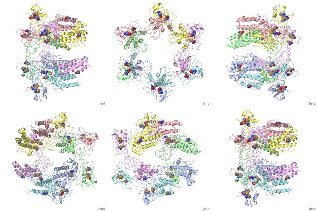
FunctionThe Flagelar hook-associated proteins FlgK (or HAP1), FlgL (or HAP3) and FliD (or HAP2) are part of the bacterial flagellum [1]. These 3 proteins seem to be located at the claw-shaped end of the flagellum and play an essential role in filament formation
|
| ||||||||||
See AlsoSee Also
- Flagella, bacterial
- Flagellar filament of bacteria
- The Bacterial Flagellar Hook
- Complete Flagellar Hook Structure determined by cryo-EM in 2016.
- Structural insights into bacterial flagellar hook similarities and specifities (2016).
3D Structures of deaminase3D Structures of deaminase
Updated on 29-November-2017
4ut1 – FlgK – Burkholderia pseudomallei
2d4x – StFlgL – Salmonella typhimurium
2d4y – StFlgK
5gna – StFlgK residues 401-467 + FliT
5h5t – StFliD D2-D3 domain
5h5v – EcFliD D1-D2-D3 domain – Escherichia coli
5h5w – EcFliD D2-D3 domain
5xlk, 5xlj – FliD D2-D3 domain – Serratia marcascens
5fhy – FliD D2-D3 domain – Pseudomonas aeruginosa
