3rmv: Difference between revisions
No edit summary |
No edit summary |
||
| Line 1: | Line 1: | ||
==Crystal Structure of Human Glycogenin-1 (GYG1) T83M mutant complexed with manganese and UDP== | ==Crystal Structure of Human Glycogenin-1 (GYG1) T83M mutant complexed with manganese and UDP== | ||
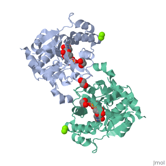
<StructureSection load='3rmv' size='340' side='right' caption='[[3rmv]], [[Resolution|resolution]] 1.82Å' scene=''> | <StructureSection load='3rmv' size='340' side='right' caption='[[3rmv]], [[Resolution|resolution]] 1.82Å' scene=''> | ||
== Structural highlights == | == Structural highlights == | ||
<table><tr><td colspan='2'>[[3rmv]] is a 1 chain structure with sequence from [http://en.wikipedia.org/wiki/ | <table><tr><td colspan='2'>[[3rmv]] is a 1 chain structure with sequence from [http://en.wikipedia.org/wiki/Human Human]. Full crystallographic information is available from [http://oca.weizmann.ac.il/oca-bin/ocashort?id=3RMV OCA]. For a <b>guided tour on the structure components</b> use [http://oca.weizmann.ac.il/oca-docs/fgij/fg.htm?mol=3RMV FirstGlance]. <br> | ||
</td></tr><tr id='ligand'><td class="sblockLbl"><b>[[Ligand|Ligands:]]</b></td><td class="sblockDat"><scene name='pdbligand=EDO:1,2-ETHANEDIOL'>EDO</scene>, <scene name='pdbligand=MG:MAGNESIUM+ION'>MG</scene>, <scene name='pdbligand=MN:MANGANESE+(II)+ION'>MN</scene>, <scene name='pdbligand=UDP:URIDINE-5-DIPHOSPHATE'>UDP</scene></td></tr> | </td></tr><tr id='ligand'><td class="sblockLbl"><b>[[Ligand|Ligands:]]</b></td><td class="sblockDat"><scene name='pdbligand=EDO:1,2-ETHANEDIOL'>EDO</scene>, <scene name='pdbligand=MG:MAGNESIUM+ION'>MG</scene>, <scene name='pdbligand=MN:MANGANESE+(II)+ION'>MN</scene>, <scene name='pdbligand=UDP:URIDINE-5-DIPHOSPHATE'>UDP</scene></td></tr> | ||
<tr id='related'><td class="sblockLbl"><b>[[Related_structure|Related:]]</b></td><td class="sblockDat">[[3qvb|3qvb]], [[3q4s|3q4s]]</td></tr> | <tr id='related'><td class="sblockLbl"><b>[[Related_structure|Related:]]</b></td><td class="sblockDat">[[3qvb|3qvb]], [[3q4s|3q4s]]</td></tr> | ||
<tr id='gene'><td class="sblockLbl"><b>[[Gene|Gene:]]</b></td><td class="sblockDat">GYG, GYG1 ([http://www.ncbi.nlm.nih.gov/Taxonomy/Browser/wwwtax.cgi?mode=Info&srchmode=5&id=9606 | <tr id='gene'><td class="sblockLbl"><b>[[Gene|Gene:]]</b></td><td class="sblockDat">GYG, GYG1 ([http://www.ncbi.nlm.nih.gov/Taxonomy/Browser/wwwtax.cgi?mode=Info&srchmode=5&id=9606 HUMAN])</td></tr> | ||
<tr id='activity'><td class="sblockLbl"><b>Activity:</b></td><td class="sblockDat"><span class='plainlinks'>[http://en.wikipedia.org/wiki/Glycogenin_glucosyltransferase Glycogenin glucosyltransferase], with EC number [http://www.brenda-enzymes.info/php/result_flat.php4?ecno=2.4.1.186 2.4.1.186] </span></td></tr> | <tr id='activity'><td class="sblockLbl"><b>Activity:</b></td><td class="sblockDat"><span class='plainlinks'>[http://en.wikipedia.org/wiki/Glycogenin_glucosyltransferase Glycogenin glucosyltransferase], with EC number [http://www.brenda-enzymes.info/php/result_flat.php4?ecno=2.4.1.186 2.4.1.186] </span></td></tr> | ||
<tr id='resources'><td class="sblockLbl"><b>Resources:</b></td><td class="sblockDat"><span class='plainlinks'>[http://oca.weizmann.ac.il/oca-docs/fgij/fg.htm?mol=3rmv FirstGlance], [http://oca.weizmann.ac.il/oca-bin/ocaids?id=3rmv OCA], [http://www.rcsb.org/pdb/explore.do?structureId=3rmv RCSB], [http://www.ebi.ac.uk/pdbsum/3rmv PDBsum]</span></td></tr> | <tr id='resources'><td class="sblockLbl"><b>Resources:</b></td><td class="sblockDat"><span class='plainlinks'>[http://oca.weizmann.ac.il/oca-docs/fgij/fg.htm?mol=3rmv FirstGlance], [http://oca.weizmann.ac.il/oca-bin/ocaids?id=3rmv OCA], [http://pdbe.org/3rmv PDBe], [http://www.rcsb.org/pdb/explore.do?structureId=3rmv RCSB], [http://www.ebi.ac.uk/pdbsum/3rmv PDBsum], [http://prosat.h-its.org/prosat/prosatexe?pdbcode=3rmv ProSAT]</span></td></tr> | ||
</table> | </table> | ||
== Disease == | |||
[[http://www.uniprot.org/uniprot/GLYG_HUMAN GLYG_HUMAN]] Glycogen storage disease due to glycogenin deficiency. The disease is caused by mutations affecting the gene represented in this entry. The disease is caused by mutations affecting the gene represented in this entry. | |||
== Function == | |||
[[http://www.uniprot.org/uniprot/GLYG_HUMAN GLYG_HUMAN]] Self-glucosylates, via an inter-subunit mechanism, to form an oligosaccharide primer that serves as substrate for glycogen synthase. | |||
<div style="background-color:#fffaf0;"> | <div style="background-color:#fffaf0;"> | ||
== Publication Abstract from PubMed == | == Publication Abstract from PubMed == | ||
| Line 17: | Line 22: | ||
From MEDLINE®/PubMed®, a database of the U.S. National Library of Medicine.<br> | From MEDLINE®/PubMed®, a database of the U.S. National Library of Medicine.<br> | ||
</div> | </div> | ||
<div class="pdbe-citations 3rmv" style="background-color:#fffaf0;"></div> | |||
==See Also== | ==See Also== | ||
| Line 25: | Line 31: | ||
</StructureSection> | </StructureSection> | ||
[[Category: Glycogenin glucosyltransferase]] | [[Category: Glycogenin glucosyltransferase]] | ||
[[Category: | [[Category: Human]] | ||
[[Category: Arrowsmith, C H]] | [[Category: Arrowsmith, C H]] | ||
[[Category: Bountra, C]] | [[Category: Bountra, C]] | ||
Revision as of 14:31, 10 December 2016
Crystal Structure of Human Glycogenin-1 (GYG1) T83M mutant complexed with manganese and UDPCrystal Structure of Human Glycogenin-1 (GYG1) T83M mutant complexed with manganese and UDP
Structural highlights
Disease[GLYG_HUMAN] Glycogen storage disease due to glycogenin deficiency. The disease is caused by mutations affecting the gene represented in this entry. The disease is caused by mutations affecting the gene represented in this entry. Function[GLYG_HUMAN] Self-glucosylates, via an inter-subunit mechanism, to form an oligosaccharide primer that serves as substrate for glycogen synthase. Publication Abstract from PubMedGlycogenin initiates the synthesis of a maltosaccharide chain covalently attached to itself on Tyr195 via a stepwise glucosylation reaction, priming glycogen synthesis. We have captured crystallographic snapshots of human glycogenin during its reaction cycle, revealing a dynamic conformational switch between ground and active states mediated by the sugar donor UDP-glucose. This switch includes the ordering of a polypeptide stretch containing Tyr195, and major movement of an approximately 30-residue "lid" segment covering the active site. The rearranged lid guides the nascent maltosaccharide chain into the active site in either an intra- or intersubunit mode dependent upon chain length and steric factors and positions the donor and acceptor sugar groups for catalysis. The Thr83Met mutation, which causes glycogen storage disease XV, is conformationally locked in the ground state and catalytically inactive. Our data highlight the conformational plasticity of glycogenin and coexistence of two modes of glucosylation as integral to its catalytic mechanism. Conformational plasticity of glycogenin and its maltosaccharide substrate during glycogen biogenesis.,Chaikuad A, Froese DS, Berridge G, von Delft F, Oppermann U, Yue WW Proc Natl Acad Sci U S A. 2011 Dec 12. PMID:22160680[1] From MEDLINE®/PubMed®, a database of the U.S. National Library of Medicine. See AlsoReferences
|
| ||||||||||||||||||||||