3pih: Difference between revisions
No edit summary |
No edit summary |
||
| Line 1: | Line 1: | ||
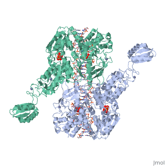
==T. maritima UvrA in complex with fluorescein-modified DNA== | ==T. maritima UvrA in complex with fluorescein-modified DNA== | ||
<StructureSection load='3pih' size='340' side='right' caption='[[3pih]], [[Resolution|resolution]] 2.90Å' scene=''> | <StructureSection load='3pih' size='340' side='right' caption='[[3pih]], [[Resolution|resolution]] 2.90Å' scene=''> | ||
== Structural highlights == | == Structural highlights == | ||
<table><tr><td colspan='2'>[[3pih]] is a 2 chain structure with sequence from [http://en.wikipedia.org/wiki/ | <table><tr><td colspan='2'>[[3pih]] is a 2 chain structure with sequence from [http://en.wikipedia.org/wiki/Atcc_43589 Atcc 43589]. Full crystallographic information is available from [http://oca.weizmann.ac.il/oca-bin/ocashort?id=3PIH OCA]. For a <b>guided tour on the structure components</b> use [http://oca.weizmann.ac.il/oca-docs/fgij/fg.htm?mol=3PIH FirstGlance]. <br> | ||
</td></tr><tr id='ligand'><td class="sblockLbl"><b>[[Ligand|Ligands:]]</b></td><td class="sblockDat"><scene name='pdbligand=PPV:PYROPHOSPHATE'>PPV</scene>, <scene name='pdbligand=ZN:ZINC+ION'>ZN</scene></td></tr> | </td></tr><tr id='ligand'><td class="sblockLbl"><b>[[Ligand|Ligands:]]</b></td><td class="sblockDat"><scene name='pdbligand=PPV:PYROPHOSPHATE'>PPV</scene>, <scene name='pdbligand=ZN:ZINC+ION'>ZN</scene></td></tr> | ||
<tr id='gene'><td class="sblockLbl"><b>[[Gene|Gene:]]</b></td><td class="sblockDat">uvrA, TM_0480 ([http://www.ncbi.nlm.nih.gov/Taxonomy/Browser/wwwtax.cgi?mode=Info&srchmode=5&id=2336 | <tr id='gene'><td class="sblockLbl"><b>[[Gene|Gene:]]</b></td><td class="sblockDat">uvrA, TM_0480 ([http://www.ncbi.nlm.nih.gov/Taxonomy/Browser/wwwtax.cgi?mode=Info&srchmode=5&id=2336 ATCC 43589])</td></tr> | ||
<tr id='resources'><td class="sblockLbl"><b>Resources:</b></td><td class="sblockDat"><span class='plainlinks'>[http://oca.weizmann.ac.il/oca-docs/fgij/fg.htm?mol=3pih FirstGlance], [http://oca.weizmann.ac.il/oca-bin/ocaids?id=3pih OCA], [http://www.rcsb.org/pdb/explore.do?structureId=3pih RCSB], [http://www.ebi.ac.uk/pdbsum/3pih PDBsum]</span></td></tr> | <tr id='resources'><td class="sblockLbl"><b>Resources:</b></td><td class="sblockDat"><span class='plainlinks'>[http://oca.weizmann.ac.il/oca-docs/fgij/fg.htm?mol=3pih FirstGlance], [http://oca.weizmann.ac.il/oca-bin/ocaids?id=3pih OCA], [http://pdbe.org/3pih PDBe], [http://www.rcsb.org/pdb/explore.do?structureId=3pih RCSB], [http://www.ebi.ac.uk/pdbsum/3pih PDBsum], [http://prosat.h-its.org/prosat/prosatexe?pdbcode=3pih ProSAT]</span></td></tr> | ||
</table> | </table> | ||
== Function == | == Function == | ||
| Line 17: | Line 18: | ||
From MEDLINE®/PubMed®, a database of the U.S. National Library of Medicine.<br> | From MEDLINE®/PubMed®, a database of the U.S. National Library of Medicine.<br> | ||
</div> | </div> | ||
<div class="pdbe-citations 3pih" style="background-color:#fffaf0;"></div> | |||
==See Also== | ==See Also== | ||
| Line 24: | Line 26: | ||
__TOC__ | __TOC__ | ||
</StructureSection> | </StructureSection> | ||
[[Category: | [[Category: Atcc 43589]] | ||
[[Category: Jaciuk, M]] | [[Category: Jaciuk, M]] | ||
[[Category: Nowak, E]] | [[Category: Nowak, E]] | ||
Revision as of 16:48, 5 August 2016
T. maritima UvrA in complex with fluorescein-modified DNAT. maritima UvrA in complex with fluorescein-modified DNA
Structural highlights
Function[UVRA_THEMA] The UvrABC repair system catalyzes the recognition and processing of DNA lesions. UvrA is an ATPase and a DNA-binding protein. A damage recognition complex composed of 2 UvrA and 2 UvrB subunits scans DNA for abnormalities. When the presence of a lesion has been verified by UvrB, the UvrA molecules dissociate (By similarity). Publication Abstract from PubMedOne of the primary pathways for removal of DNA damage is nucleotide excision repair (NER). In bacteria, the UvrA protein is the component of NER that locates the lesion. A notable feature of NER is its ability to act on many DNA modifications that vary in chemical structure. So far, the mechanism underlying this broad specificity has been unclear. Here, we report the first crystal structure of a UvrA protein in complex with a chemically modified oligonucleotide. The structure shows that the UvrA dimer does not contact the site of lesion directly, but rather binds the DNA regions on both sides of the modification. The DNA region harboring the modification is deformed, with the double helix bent and unwound. UvrA uses damage-induced deformations of the DNA and a less rigid structure of the modified double helix for indirect readout of the lesion. Structure of UvrA nucleotide excision repair protein in complex with modified DNA.,Jaciuk M, Nowak E, Skowronek K, Tanska A, Nowotny M Nat Struct Mol Biol. 2011 Feb;18(2):191-7. Epub 2011 Jan 16. PMID:21240268[1] From MEDLINE®/PubMed®, a database of the U.S. National Library of Medicine. See AlsoReferences
|
| ||||||||||||||||||