Myoglobin: Difference between revisions
Michal Harel (talk | contribs) No edit summary |
Michal Harel (talk | contribs) No edit summary |
||
| Line 37: | Line 37: | ||
**[[1mbs]] – Mb - seal<br /> | **[[1mbs]] – Mb - seal<br /> | ||
**[[1uvy]] – Mb – ''Paramecium caudatum''<br /> | **[[1uvy]] – Mb – ''Paramecium caudatum''<br /> | ||
**[[3qm5]], [[3qm6]] – btMb – blackfin tuna | |||
* '''Mb mutants uncomplexed''' | * '''Mb mutants uncomplexed''' | ||
Revision as of 13:12, 3 May 2016
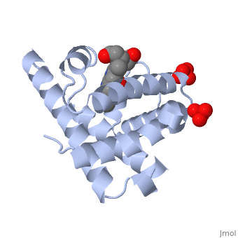
Myoglobin is a globular protein whose function is to store molecular oxygen in muscles (myo = muscles)[1]. It has two main components: a single polypeptide chain, and heme ligand. The heme ligand is only to the surface; the majority of it is buried inside the protein. The overall of myoglobin is approximately disc-shaped with a diameter that is about twice its thickness. The overall fold of the protein is conserved, especially the core of the protein (shown in purple), but the sequence is more on the surface. The globin consists mostly of alpha helices shown in ; it has no beta sheets and its non-helical segments mostly serve as links that connect the helices. Look down the barrel of some of the longer helices. Are they all straight? The eight structurally conserved alpha helices are labelled . The protein is colored as a N-->C rainbow in this view; the N terminus is blue, while the C terminus is red. The , and specifically the iron atom in the middle of the heme, is what binds oxygen in myoglobin. In this representation, the heme alone is shown in ball and stick form with its C, N and O atoms displayed as grey, blue, and red balls respectively. The iron atom is shown in orange, and is in spacefilling mode to better illustrate its interactions with the heme. The iron is bound by four nitrogen atoms found in the heme ring, as well as an from the protein chain. Which amino acid from the myoglobin protein binds to the iron? Notice that in the oxygenated state, the iron is in the plane of the heme ring. In the (no oxygen) state, the Fe atom is slightly above the plane of the heme, and a second coordinates with the iron in the heme ring. For myoglobin complex with O2 see Oxymyoglobin For porphyrin see Porphyrin. See also Molecular Playground/Myoglobin, Extremophile and Extremophiles.
Myoglobin-Physeter-catodon-structure (Spanish)
|
| ||||||||||

3D Structures of Myoglobin3D Structures of Myoglobin
Updated on 03-May-2016
Myoglobin (Mb) is an oxygen binding protein found in muscle tissue. It contains a heme group. Metmyoglobin (MMb) is the oxidized form of myoglobin.
External ResourcesExternal Resources
ReferencesReferences
- ↑ Ordway GA, Garry DJ. Myoglobin: an essential hemoprotein in striated muscle. J Exp Biol. 2004 Sep;207(Pt 20):3441-6. PMID:15339940 doi:http://dx.doi.org/10.1242/jeb.01172