1e9z: Difference between revisions
No edit summary |
No edit summary |
||
| Line 4: | Line 4: | ||
|PDB= 1e9z |SIZE=350|CAPTION= <scene name='initialview01'>1e9z</scene>, resolution 3.0Å | |PDB= 1e9z |SIZE=350|CAPTION= <scene name='initialview01'>1e9z</scene>, resolution 3.0Å | ||
|SITE= <scene name='pdbsite=NI1:Ni+Binding+Site+For+Residue+B2002'>NI1</scene> | |SITE= <scene name='pdbsite=NI1:Ni+Binding+Site+For+Residue+B2002'>NI1</scene> | ||
|LIGAND= <scene name='pdbligand=NI:NICKEL (II) ION'>NI</scene> | |LIGAND= <scene name='pdbligand=KCX:LYSINE+NZ-CARBOXYLIC+ACID'>KCX</scene>, <scene name='pdbligand=NI:NICKEL+(II)+ION'>NI</scene> | ||
|ACTIVITY= [http://en.wikipedia.org/wiki/Urease Urease], with EC number [http://www.brenda-enzymes.info/php/result_flat.php4?ecno=3.5.1.5 3.5.1.5] | |ACTIVITY= <span class='plainlinks'>[http://en.wikipedia.org/wiki/Urease Urease], with EC number [http://www.brenda-enzymes.info/php/result_flat.php4?ecno=3.5.1.5 3.5.1.5] </span> | ||
|GENE= | |GENE= | ||
|DOMAIN= | |||
|RELATEDENTRY= | |||
|RESOURCES=<span class='plainlinks'>[http://oca.weizmann.ac.il/oca-docs/fgij/fg.htm?mol=1e9z FirstGlance], [http://oca.weizmann.ac.il/oca-bin/ocaids?id=1e9z OCA], [http://www.ebi.ac.uk/pdbsum/1e9z PDBsum], [http://www.rcsb.org/pdb/explore.do?structureId=1e9z RCSB]</span> | |||
}} | }} | ||
| Line 26: | Line 29: | ||
[[Category: Oh, B H.]] | [[Category: Oh, B H.]] | ||
[[Category: Oh, S T.]] | [[Category: Oh, S T.]] | ||
[[Category: dodecamer]] | [[Category: dodecamer]] | ||
[[Category: helicobacter]] | [[Category: helicobacter]] | ||
[[Category: urease]] | [[Category: urease]] | ||
''Page seeded by [http://oca.weizmann.ac.il/oca OCA ] on | ''Page seeded by [http://oca.weizmann.ac.il/oca OCA ] on Sun Mar 30 19:58:19 2008'' | ||
Revision as of 19:58, 30 March 2008
| |||||||
| , resolution 3.0Å | |||||||
|---|---|---|---|---|---|---|---|
| Sites: | |||||||
| Ligands: | , | ||||||
| Activity: | Urease, with EC number 3.5.1.5 | ||||||
| Resources: | FirstGlance, OCA, PDBsum, RCSB | ||||||
| Coordinates: | save as pdb, mmCIF, xml | ||||||
CRYSTAL STRUCTURE OF HELICOBACTER PYLORI UREASE
OverviewOverview
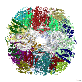
Helicobacter pylori, an etiologic agent in a variety of gastroduodenal diseases, produces a large amount of urease, which is believed to neutralize gastric acid by producing ammonia for the survival of the bacteria. Up to 30% of the enzyme associates with the surface of intact cells upon lysis of neighboring bacteria. The role of the enzyme at the extracellular location has been a subject of controversy because the purified enzyme is irreversibly inactivated below pH 5. We have determined the crystal structure of H. pylori urease, which has a 1.1 MDa spherical assembly of 12 catalytic units with an outer diameter of approximately 160 A. Under physiologically relevant conditions, the activity of the enzyme remains unaffected down to pH 3. Activity assays under different conditions indicated that the cluster of the 12 active sites on the supramolecular assembly may be critical for the survival of the enzyme at low pH. The structure provides a novel example of a molecular assembly adapted for acid resistance that, together with the low Km value of the enzyme, is likely to enable the organism to inhabit the hostile niche.
About this StructureAbout this Structure
1E9Z is a Protein complex structure of sequences from Helicobacter pylori. Full crystallographic information is available from OCA.
ReferenceReference
Supramolecular assembly and acid resistance of Helicobacter pylori urease., Ha NC, Oh ST, Sung JY, Cha KA, Lee MH, Oh BH, Nat Struct Biol. 2001 Jun;8(6):505-9. PMID:11373617
Page seeded by OCA on Sun Mar 30 19:58:19 2008