1e2h: Difference between revisions
No edit summary |
No edit summary |
||
| Line 4: | Line 4: | ||
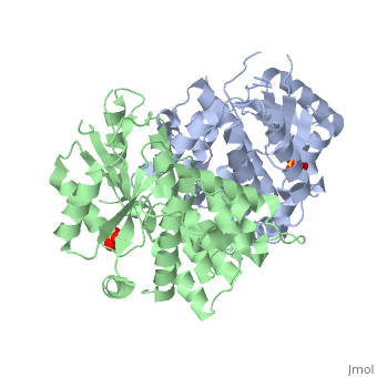
|PDB= 1e2h |SIZE=350|CAPTION= <scene name='initialview01'>1e2h</scene>, resolution 1.9Å | |PDB= 1e2h |SIZE=350|CAPTION= <scene name='initialview01'>1e2h</scene>, resolution 1.9Å | ||
|SITE= | |SITE= | ||
|LIGAND= <scene name='pdbligand=SO4:SULFATE ION'>SO4</scene> | |LIGAND= <scene name='pdbligand=SO4:SULFATE+ION'>SO4</scene> | ||
|ACTIVITY= [http://en.wikipedia.org/wiki/Thymidine_kinase Thymidine kinase], with EC number [http://www.brenda-enzymes.info/php/result_flat.php4?ecno=2.7.1.21 2.7.1.21] | |ACTIVITY= <span class='plainlinks'>[http://en.wikipedia.org/wiki/Thymidine_kinase Thymidine kinase], with EC number [http://www.brenda-enzymes.info/php/result_flat.php4?ecno=2.7.1.21 2.7.1.21] </span> | ||
|GENE= | |GENE= | ||
|DOMAIN= | |||
|RELATEDENTRY= | |||
|RESOURCES=<span class='plainlinks'>[http://oca.weizmann.ac.il/oca-docs/fgij/fg.htm?mol=1e2h FirstGlance], [http://oca.weizmann.ac.il/oca-bin/ocaids?id=1e2h OCA], [http://www.ebi.ac.uk/pdbsum/1e2h PDBsum], [http://www.rcsb.org/pdb/explore.do?structureId=1e2h RCSB]</span> | |||
}} | }} | ||
| Line 16: | Line 19: | ||
==About this Structure== | ==About this Structure== | ||
1E2H is a [[Single protein]] structure of sequence from [http://en.wikipedia.org/wiki/ | 1E2H is a [[Single protein]] structure of sequence from [http://en.wikipedia.org/wiki/Human_herpesvirus_1 Human herpesvirus 1]. Full crystallographic information is available from [http://oca.weizmann.ac.il/oca-bin/ocashort?id=1E2H OCA]. | ||
==Reference== | ==Reference== | ||
Nucleoside binding site of herpes simplex type 1 thymidine kinase analyzed by X-ray crystallography., Vogt J, Perozzo R, Pautsch A, Prota A, Schelling P, Pilger B, Folkers G, Scapozza L, Schulz GE, Proteins. 2000 Dec 1;41(4):545-53. PMID:[http://www.ncbi.nlm.nih.gov/pubmed/11056041 11056041] | Nucleoside binding site of herpes simplex type 1 thymidine kinase analyzed by X-ray crystallography., Vogt J, Perozzo R, Pautsch A, Prota A, Schelling P, Pilger B, Folkers G, Scapozza L, Schulz GE, Proteins. 2000 Dec 1;41(4):545-53. PMID:[http://www.ncbi.nlm.nih.gov/pubmed/11056041 11056041] | ||
[[Category: Human herpesvirus | [[Category: Human herpesvirus 1]] | ||
[[Category: Single protein]] | [[Category: Single protein]] | ||
[[Category: Thymidine kinase]] | [[Category: Thymidine kinase]] | ||
| Line 26: | Line 29: | ||
[[Category: Schulz, G E.]] | [[Category: Schulz, G E.]] | ||
[[Category: Vogt, J.]] | [[Category: Vogt, J.]] | ||
[[Category: adenine analog]] | [[Category: adenine analog]] | ||
[[Category: enzyme-prodrug gene therapy]] | [[Category: enzyme-prodrug gene therapy]] | ||
| Line 33: | Line 35: | ||
[[Category: x-ray crystallography]] | [[Category: x-ray crystallography]] | ||
''Page seeded by [http://oca.weizmann.ac.il/oca OCA ] on | ''Page seeded by [http://oca.weizmann.ac.il/oca OCA ] on Sun Mar 30 19:53:39 2008'' | ||
Revision as of 19:53, 30 March 2008
| |||||||
| , resolution 1.9Å | |||||||
|---|---|---|---|---|---|---|---|
| Ligands: | |||||||
| Activity: | Thymidine kinase, with EC number 2.7.1.21 | ||||||
| Resources: | FirstGlance, OCA, PDBsum, RCSB | ||||||
| Coordinates: | save as pdb, mmCIF, xml | ||||||
THE NUCLEOSIDE BINDING SITE OF HERPES SIMPLEX TYPE 1 THYMIDINE KINASE ANALYZED BY X-RAY CRYSTALLOGRAPHY
OverviewOverview
The crystal structures of the full-length Herpes simplex virus type 1 thymidine kinase in its unligated form and in a complex with an adenine analogue have been determined at 1.9 A resolution. The unligated enzyme contains four water molecules in the thymidine pocket and reveals a small induced fit on substrate binding. The structure of the ligated enzyme shows for the first time a bound adenine analogue after numerous complexes with thymine and guanine analogues have been reported. The adenine analogue constitutes a new lead compound for enzyme-prodrug gene therapy. In addition, the structure of mutant Q125N modifying the binding site of the natural substrate thymidine in complex with this substrate has been established at 2.5 A resolution. It reveals that neither the binding mode of thymidine nor the polypeptide backbone conformation is altered, except that the two major hydrogen bonds to thymidine are replaced by a single water-mediated hydrogen bond, which improves the relative acceptance of the prodrugs aciclovir and ganciclovir compared with the natural substrate. Accordingly, the mutant structure represents a first step toward improving the virus-directed enzyme-prodrug gene therapy by enzyme engineering.
About this StructureAbout this Structure
1E2H is a Single protein structure of sequence from Human herpesvirus 1. Full crystallographic information is available from OCA.
ReferenceReference
Nucleoside binding site of herpes simplex type 1 thymidine kinase analyzed by X-ray crystallography., Vogt J, Perozzo R, Pautsch A, Prota A, Schelling P, Pilger B, Folkers G, Scapozza L, Schulz GE, Proteins. 2000 Dec 1;41(4):545-53. PMID:11056041
Page seeded by OCA on Sun Mar 30 19:53:39 2008