2pbd: Difference between revisions
No edit summary |
No edit summary |
||
| Line 4: | Line 4: | ||
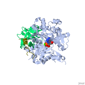
|PDB= 2pbd |SIZE=350|CAPTION= <scene name='initialview01'>2pbd</scene>, resolution 1.501Å | |PDB= 2pbd |SIZE=350|CAPTION= <scene name='initialview01'>2pbd</scene>, resolution 1.501Å | ||
|SITE= | |SITE= | ||
|LIGAND= <scene name='pdbligand=CA:CALCIUM+ION'>CA</scene> and <scene name='pdbligand=ATP:ADENOSINE-5 | |LIGAND= <scene name='pdbligand=CA:CALCIUM+ION'>CA</scene> and <scene name='pdbligand=ATP:ADENOSINE-5'-TRIPHOSPHATE'>ATP</scene> | ||
|ACTIVITY= | |ACTIVITY= | ||
|GENE= PFN1 ([http://www.ncbi.nlm.nih.gov/Taxonomy/Browser/wwwtax.cgi?mode=Info&srchmode=5&id=9606 Homo sapiens]) | |GENE= PFN1 ([http://www.ncbi.nlm.nih.gov/Taxonomy/Browser/wwwtax.cgi?mode=Info&srchmode=5&id=9606 Homo sapiens]) | ||
| Line 31: | Line 31: | ||
[[Category: ternary complex; profilin; actin; vasp; poly-proline; loading poly-pro site; gab domain]] | [[Category: ternary complex; profilin; actin; vasp; poly-proline; loading poly-pro site; gab domain]] | ||
''Page seeded by [http://oca.weizmann.ac.il/oca OCA ] on | ''Page seeded by [http://oca.weizmann.ac.il/oca OCA ] on Sun Mar 23 15:38:08 2008'' | ||
Revision as of 16:38, 23 March 2008
| |||||||
| , resolution 1.501Å | |||||||
|---|---|---|---|---|---|---|---|
| Ligands: | and | ||||||
| Gene: | PFN1 (Homo sapiens) | ||||||
| Coordinates: | save as pdb, mmCIF, xml | ||||||
Ternary complex of profilin-actin with the poly-PRO-GAB domain of VASP*
OverviewOverview
Cells sustain high rates of actin filament elongation by maintaining a large pool of actin monomers above the critical concentration for polymerization. Profilin-actin complexes constitute the largest fraction of polymerization-competent actin monomers. Filament elongation factors such as Ena/VASP and formin catalyze the transition of profilin-actin from the cellular pool onto the barbed end of growing filaments. The molecular bases of this process are poorly understood. Here we present structural and energetic evidence for two consecutive steps of the elongation mechanism: the recruitment of profilin-actin by the last poly-Pro segment of vasodilator-stimulated phosphoprotein (VASP) and the binding of profilin-actin simultaneously to this poly-Pro and to the G-actin-binding (GAB) domain of VASP. The actin monomer bound at the GAB domain is proposed to be in position to join the barbed end of the growing filament concurrently with the release of profilin.
About this StructureAbout this Structure
2PBD is a Protein complex structure of sequences from Homo sapiens and Oryctolagus cuniculus. Full crystallographic information is available from OCA.
ReferenceReference
Structural basis for the recruitment of profilin-actin complexes during filament elongation by Ena/VASP., Ferron F, Rebowski G, Lee SH, Dominguez R, EMBO J. 2007 Oct 31;26(21):4597-606. Epub 2007 Oct 4. PMID:17914456
Page seeded by OCA on Sun Mar 23 15:38:08 2008