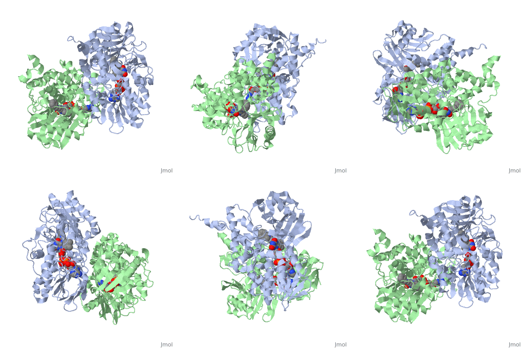
1gos: Difference between revisions
No edit summary |
No edit summary |
||
| Line 2: | Line 2: | ||
<StructureSection load='1gos' size='340' side='right' caption='[[1gos]], [[Resolution|resolution]] 3.00Å' scene=''> | <StructureSection load='1gos' size='340' side='right' caption='[[1gos]], [[Resolution|resolution]] 3.00Å' scene=''> | ||
== Structural highlights == | == Structural highlights == | ||
<table><tr><td colspan='2'>[[1gos]] is a 2 chain structure with sequence from [http://en.wikipedia.org/wiki/ | <table><tr><td colspan='2'>[[1gos]] is a 2 chain structure with sequence from [http://en.wikipedia.org/wiki/Human Human]. Full crystallographic information is available from [http://oca.weizmann.ac.il/oca-bin/ocashort?id=1GOS OCA]. For a <b>guided tour on the structure components</b> use [http://oca.weizmann.ac.il/oca-docs/fgij/fg.htm?mol=1GOS FirstGlance]. <br> | ||
</td></tr><tr id='ligand'><td class="sblockLbl"><b>[[Ligand|Ligands:]]</b></td><td class="sblockDat"><scene name='pdbligand=FAD:FLAVIN-ADENINE+DINUCLEOTIDE'>FAD</scene>, <scene name='pdbligand=NYP:N-[(E)-METHYL](PHENYL)-N-[(E)-2-PROPENYLIDENE]METHANAMINIUM'>NYP</scene></td></tr> | </td></tr><tr id='ligand'><td class="sblockLbl"><b>[[Ligand|Ligands:]]</b></td><td class="sblockDat"><scene name='pdbligand=FAD:FLAVIN-ADENINE+DINUCLEOTIDE'>FAD</scene>, <scene name='pdbligand=NYP:N-[(E)-METHYL](PHENYL)-N-[(E)-2-PROPENYLIDENE]METHANAMINIUM'>NYP</scene></td></tr> | ||
<tr id='related'><td class="sblockLbl"><b>[[Related_structure|Related:]]</b></td><td class="sblockDat">[[1h8r|1h8r]]</td></tr> | <tr id='related'><td class="sblockLbl"><b>[[Related_structure|Related:]]</b></td><td class="sblockDat">[[1h8r|1h8r]]</td></tr> | ||
<tr id='activity'><td class="sblockLbl"><b>Activity:</b></td><td class="sblockDat"><span class='plainlinks'>[http://en.wikipedia.org/wiki/Monoamine_oxidase Monoamine oxidase], with EC number [http://www.brenda-enzymes.info/php/result_flat.php4?ecno=1.4.3.4 1.4.3.4] </span></td></tr> | <tr id='activity'><td class="sblockLbl"><b>Activity:</b></td><td class="sblockDat"><span class='plainlinks'>[http://en.wikipedia.org/wiki/Monoamine_oxidase Monoamine oxidase], with EC number [http://www.brenda-enzymes.info/php/result_flat.php4?ecno=1.4.3.4 1.4.3.4] </span></td></tr> | ||
<tr id='resources'><td class="sblockLbl"><b>Resources:</b></td><td class="sblockDat"><span class='plainlinks'>[http://oca.weizmann.ac.il/oca-docs/fgij/fg.htm?mol=1gos FirstGlance], [http://oca.weizmann.ac.il/oca-bin/ocaids?id=1gos OCA], [http://www.rcsb.org/pdb/explore.do?structureId=1gos RCSB], [http://www.ebi.ac.uk/pdbsum/1gos PDBsum]</span></td></tr> | <tr id='resources'><td class="sblockLbl"><b>Resources:</b></td><td class="sblockDat"><span class='plainlinks'>[http://oca.weizmann.ac.il/oca-docs/fgij/fg.htm?mol=1gos FirstGlance], [http://oca.weizmann.ac.il/oca-bin/ocaids?id=1gos OCA], [http://pdbe.org/1gos PDBe], [http://www.rcsb.org/pdb/explore.do?structureId=1gos RCSB], [http://www.ebi.ac.uk/pdbsum/1gos PDBsum]</span></td></tr> | ||
</table> | </table> | ||
== Evolutionary Conservation == | == Evolutionary Conservation == | ||
| Line 26: | Line 26: | ||
From MEDLINE®/PubMed®, a database of the U.S. National Library of Medicine.<br> | From MEDLINE®/PubMed®, a database of the U.S. National Library of Medicine.<br> | ||
</div> | </div> | ||
<div class="pdbe-citations 1gos" style="background-color:#fffaf0;"></div> | |||
==See Also== | ==See Also== | ||
| Line 34: | Line 35: | ||
__TOC__ | __TOC__ | ||
</StructureSection> | </StructureSection> | ||
[[Category: | [[Category: Human]] | ||
[[Category: Monoamine oxidase]] | [[Category: Monoamine oxidase]] | ||
[[Category: Binda, C]] | [[Category: Binda, C]] | ||
Revision as of 09:09, 10 September 2015
HUMAN MONOAMINE OXIDASE BHUMAN MONOAMINE OXIDASE B
Structural highlights
Evolutionary Conservation Check, as determined by ConSurfDB. You may read the explanation of the method and the full data available from ConSurf. Publication Abstract from PubMedMonoamine oxidase B (MAO B) is a mitochondrial outermembrane flavoenzyme that is a well-known target for antidepressant and neuroprotective drugs. We determined the structure of the human enzyme to 3 A resolution. The enzyme binds to the membrane through a C-terminal transmembrane helix and apolar loops located at various positions in the sequence. The electron density shows that pargyline, an analog of the clinically used MAO B inhibitor, deprenyl, binds covalently to the flavin N5 atom. The active site of MAO B consists of a 420 A(3)-hydrophobic substrate cavity interconnected to an entrance cavity of 290 A(3). The recognition site for the substrate amino group is an aromatic cage formed by Tyr 398 and Tyr 435. The structure provides a framework for probing the catalytic mechanism, understanding the differences between the B- and A-monoamine oxidase isoforms and designing specific inhibitors. Structure of human monoamine oxidase B, a drug target for the treatment of neurological disorders.,Binda C, Newton-Vinson P, Hubalek F, Edmondson DE, Mattevi A Nat Struct Biol. 2002 Jan;9(1):22-6. PMID:11753429[1] From MEDLINE®/PubMed®, a database of the U.S. National Library of Medicine. See AlsoReferences
|
| ||||||||||||||||||||
