1a0i: Difference between revisions
No edit summary |
No edit summary |
||
| Line 8: | Line 8: | ||
<tr id='resources'><td class="sblockLbl"><b>Resources:</b></td><td class="sblockDat"><span class='plainlinks'>[http://oca.weizmann.ac.il/oca-docs/fgij/fg.htm?mol=1a0i FirstGlance], [http://oca.weizmann.ac.il/oca-bin/ocaids?id=1a0i OCA], [http://www.rcsb.org/pdb/explore.do?structureId=1a0i RCSB], [http://www.ebi.ac.uk/pdbsum/1a0i PDBsum]</span></td></tr> | <tr id='resources'><td class="sblockLbl"><b>Resources:</b></td><td class="sblockDat"><span class='plainlinks'>[http://oca.weizmann.ac.il/oca-docs/fgij/fg.htm?mol=1a0i FirstGlance], [http://oca.weizmann.ac.il/oca-bin/ocaids?id=1a0i OCA], [http://www.rcsb.org/pdb/explore.do?structureId=1a0i RCSB], [http://www.ebi.ac.uk/pdbsum/1a0i PDBsum]</span></td></tr> | ||
</table> | </table> | ||
== Function == | |||
[[http://www.uniprot.org/uniprot/DNLI_BPT7 DNLI_BPT7]] DNA ligase, which is expressed in the early stage of lytic development, has been implicated in T7 DNA synthesis and genetic recombination. It may also play a role in T7 DNA repair. | |||
== Evolutionary Conservation == | == Evolutionary Conservation == | ||
[[Image:Consurf_key_small.gif|200px|right]] | [[Image:Consurf_key_small.gif|200px|right]] | ||
Revision as of 00:38, 26 December 2014
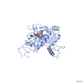
ATP-DEPENDENT DNA LIGASE FROM BACTERIOPHAGE T7 COMPLEX WITH ATPATP-DEPENDENT DNA LIGASE FROM BACTERIOPHAGE T7 COMPLEX WITH ATP
Structural highlights
Function[DNLI_BPT7] DNA ligase, which is expressed in the early stage of lytic development, has been implicated in T7 DNA synthesis and genetic recombination. It may also play a role in T7 DNA repair. Evolutionary Conservation Check, as determined by ConSurfDB. You may read the explanation of the method and the full data available from ConSurf. Publication Abstract from PubMedThe crystal structure of the ATP-dependent DNA ligase from bacteriophage T7 has been solved at 2.6 A resolution. The protein comprises two domains with a deep cleft running between them. The structure of a complex with ATP reveals that the nucleotide binding pocket is situated on the larger N-terminal domain, at the base of the cleft between the two domains of the enzyme. Comparison of the overall domain structure with that of DNA methyltransferases, coupled with other evidence, suggests that DNA also binds in this cleft. Since this structure is the first of the nucleotidyltransferase superfamily, which includes the eukaryotic mRNA capping enzymes, the relationship between the structure of DNA ligase and that of other nucleotidyltransferases is also discussed. Crystal structure of an ATP-dependent DNA ligase from bacteriophage T7.,Subramanya HS, Doherty AJ, Ashford SR, Wigley DB Cell. 1996 May 17;85(4):607-15. PMID:8653795[1] From MEDLINE®/PubMed®, a database of the U.S. National Library of Medicine. See Also
References |
| ||||||||||||||||||||