2djh: Difference between revisions
No edit summary |
No edit summary |
||
| Line 1: | Line 1: | ||
[[Image:2djh.gif|left|200px]] | [[Image:2djh.gif|left|200px]] | ||
'''Crystal structure of the carboxy-terminal ribonuclease domain of Colicin E5''' | {{Structure | ||
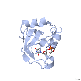
|PDB= 2djh |SIZE=350|CAPTION= <scene name='initialview01'>2djh</scene>, resolution 1.90Å | |||
|SITE= | |||
|LIGAND= <scene name='pdbligand=3PD:2-AMINO-9-(2-DEOXY-3-O-PHOSPHONOPENTOFURANOSYL)-1,9-DIHYDRO-6H-PURIN-6-ONE'>3PD</scene> | |||
|ACTIVITY= | |||
|GENE= | |||
}} | |||
'''Crystal structure of the carboxy-terminal ribonuclease domain of Colicin E5''' | |||
==Overview== | ==Overview== | ||
| Line 7: | Line 16: | ||
==About this Structure== | ==About this Structure== | ||
2DJH is a [ | 2DJH is a [[Single protein]] structure of sequence from [http://en.wikipedia.org/wiki/Escherichia_coli Escherichia coli]. Full crystallographic information is available from [http://oca.weizmann.ac.il/oca-bin/ocashort?id=2DJH OCA]. | ||
==Reference== | ==Reference== | ||
Structural basis for sequence-dependent recognition of colicin E5 tRNase by mimicking the mRNA-tRNA interaction., Yajima S, Inoue S, Ogawa T, Nonaka T, Ohsawa K, Masaki H, Nucleic Acids Res. 2006;34(21):6074-82. Epub 2006 Nov 11. PMID:[http:// | Structural basis for sequence-dependent recognition of colicin E5 tRNase by mimicking the mRNA-tRNA interaction., Yajima S, Inoue S, Ogawa T, Nonaka T, Ohsawa K, Masaki H, Nucleic Acids Res. 2006;34(21):6074-82. Epub 2006 Nov 11. PMID:[http://www.ncbi.nlm.nih.gov/pubmed/17099236 17099236] | ||
[[Category: Escherichia coli]] | [[Category: Escherichia coli]] | ||
[[Category: Single protein]] | [[Category: Single protein]] | ||
| Line 22: | Line 31: | ||
[[Category: alpha/beta protein]] | [[Category: alpha/beta protein]] | ||
''Page seeded by [http://oca.weizmann.ac.il/oca OCA ] on Thu | ''Page seeded by [http://oca.weizmann.ac.il/oca OCA ] on Thu Mar 20 16:27:56 2008'' | ||
Revision as of 17:27, 20 March 2008
| |||||||
| , resolution 1.90Å | |||||||
|---|---|---|---|---|---|---|---|
| Ligands: | |||||||
| Coordinates: | save as pdb, mmCIF, xml | ||||||
Crystal structure of the carboxy-terminal ribonuclease domain of Colicin E5
OverviewOverview
Colicin E5--a tRNase toxin--specifically cleaves QUN (Q: queuosine) anticodons of the Escherichia coli tRNAs for Tyr, His, Asn and Asp. Here, we report the crystal structure of the C-terminal ribonuclease domain (CRD) of E5 complexed with a substrate analog, namely, dGpdUp, at a resolution of 1.9 A. Thisstructure is the first to reveal the substrate recognition mechanism of sequence-specific ribonucleases. E5-CRD realized the strict recognition for both the guanine and uracil bases of dGpdUp forming Watson-Crick-type hydrogen bonds and ring stacking interactions, thus mimicking the codons of mRNAs to bind to tRNA anticodons. The docking model of E5-CRD with tRNA also suggests its substrate preference for tRNA over ssRNA. In addition, the structure of E5-CRD/dGpdUp along with the mutational analysis suggests that Arg33 may play an important role in the catalytic activity, and Lys25/Lys60 may also be involved without His in E5-CRD. Finally, the comparison of the structures of E5-CRD/dGpdUp and E5-CRD/ImmE5 (an inhibitor protein) complexes suggests that the binding mode of E5-CRD and ImmE5 mimics that of mRNA and tRNA; this may represent the evolutionary pathway of these proteins from the RNA-RNA interaction through the RNA-protein interaction of tRNA/E5-CRD.
About this StructureAbout this Structure
2DJH is a Single protein structure of sequence from Escherichia coli. Full crystallographic information is available from OCA.
ReferenceReference
Structural basis for sequence-dependent recognition of colicin E5 tRNase by mimicking the mRNA-tRNA interaction., Yajima S, Inoue S, Ogawa T, Nonaka T, Ohsawa K, Masaki H, Nucleic Acids Res. 2006;34(21):6074-82. Epub 2006 Nov 11. PMID:17099236
Page seeded by OCA on Thu Mar 20 16:27:56 2008