1a3n: Difference between revisions
No edit summary |
No edit summary |
||
| Line 3: | Line 3: | ||
== Structural highlights == | == Structural highlights == | ||
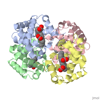
<table><tr><td colspan='2'>[[1a3n]] is a 4 chain structure with sequence from [http://en.wikipedia.org/wiki/Homo_sapiens Homo sapiens]. Full crystallographic information is available from [http://oca.weizmann.ac.il/oca-bin/ocashort?id=1A3N OCA]. For a <b>guided tour on the structure components</b> use [http://oca.weizmann.ac.il/oca-docs/fgij/fg.htm?mol=1A3N FirstGlance]. <br> | <table><tr><td colspan='2'>[[1a3n]] is a 4 chain structure with sequence from [http://en.wikipedia.org/wiki/Homo_sapiens Homo sapiens]. Full crystallographic information is available from [http://oca.weizmann.ac.il/oca-bin/ocashort?id=1A3N OCA]. For a <b>guided tour on the structure components</b> use [http://oca.weizmann.ac.il/oca-docs/fgij/fg.htm?mol=1A3N FirstGlance]. <br> | ||
</td></tr><tr><td class="sblockLbl"><b>[[Ligand|Ligands:]]</b></td><td class="sblockDat"><scene name='pdbligand=HEM:PROTOPORPHYRIN+IX+CONTAINING+FE'>HEM</scene>< | </td></tr><tr id='ligand'><td class="sblockLbl"><b>[[Ligand|Ligands:]]</b></td><td class="sblockDat"><scene name='pdbligand=HEM:PROTOPORPHYRIN+IX+CONTAINING+FE'>HEM</scene></td></tr> | ||
<tr><td class="sblockLbl"><b>Resources:</b></td><td class="sblockDat"><span class='plainlinks'>[http://oca.weizmann.ac.il/oca-docs/fgij/fg.htm?mol=1a3n FirstGlance], [http://oca.weizmann.ac.il/oca-bin/ocaids?id=1a3n OCA], [http://www.rcsb.org/pdb/explore.do?structureId=1a3n RCSB], [http://www.ebi.ac.uk/pdbsum/1a3n PDBsum]</span></td></tr> | <tr id='resources'><td class="sblockLbl"><b>Resources:</b></td><td class="sblockDat"><span class='plainlinks'>[http://oca.weizmann.ac.il/oca-docs/fgij/fg.htm?mol=1a3n FirstGlance], [http://oca.weizmann.ac.il/oca-bin/ocaids?id=1a3n OCA], [http://www.rcsb.org/pdb/explore.do?structureId=1a3n RCSB], [http://www.ebi.ac.uk/pdbsum/1a3n PDBsum]</span></td></tr> | ||
<table> | </table> | ||
== Disease == | == Disease == | ||
[[http://www.uniprot.org/uniprot/HBB_HUMAN HBB_HUMAN]] Defects in HBB may be a cause of Heinz body anemias (HEIBAN) [MIM:[http://omim.org/entry/140700 140700]]. This is a form of non-spherocytic hemolytic anemia of Dacie type 1. After splenectomy, which has little benefit, basophilic inclusions called Heinz bodies are demonstrable in the erythrocytes. Before splenectomy, diffuse or punctate basophilia may be evident. Most of these cases are probably instances of hemoglobinopathy. The hemoglobin demonstrates heat lability. Heinz bodies are observed also with the Ivemark syndrome (asplenia with cardiovascular anomalies) and with glutathione peroxidase deficiency.<ref>PMID:186485</ref> <ref>PMID:6259091</ref> <ref>PMID:2599881</ref> <ref>PMID:8704193</ref> Defects in HBB are the cause of beta-thalassemia (B-THAL) [MIM:[http://omim.org/entry/613985 613985]]. A form of thalassemia. Thalassemias are common monogenic diseases occurring mostly in Mediterranean and Southeast Asian populations. The hallmark of beta-thalassemia is an imbalance in globin-chain production in the adult HbA molecule. Absence of beta chain causes beta(0)-thalassemia, while reduced amounts of detectable beta globin causes beta(+)-thalassemia. In the severe forms of beta-thalassemia, the excess alpha globin chains accumulate in the developing erythroid precursors in the marrow. Their deposition leads to a vast increase in erythroid apoptosis that in turn causes ineffective erythropoiesis and severe microcytic hypochromic anemia. Clinically, beta-thalassemia is divided into thalassemia major which is transfusion dependent, thalassemia intermedia (of intermediate severity), and thalassemia minor that is asymptomatic.<ref>PMID:1971109</ref> Defects in HBB are the cause of sickle cell anemia (SKCA) [MIM:[http://omim.org/entry/603903 603903]]; also known as sickle cell disease. Sickle cell anemia is characterized by abnormally shaped red cells resulting in chronic anemia and periodic episodes of pain, serious infections and damage to vital organs. Normal red blood cells are round and flexible and flow easily through blood vessels, but in sickle cell anemia, the abnormal hemoglobin (called Hb S) causes red blood cells to become stiff. They are C-shaped and resembles a sickle. These stiffer red blood cells can led to microvascular occlusion thus cutting off the blood supply to nearby tissues. Defects in HBB are the cause of beta-thalassemia dominant inclusion body type (B-THALIB) [MIM:[http://omim.org/entry/603902 603902]]. An autosomal dominant form of beta thalassemia characterized by moderate anemia, lifelong jaundice, cholelithiasis and splenomegaly, marked morphologic changes in the red cells, erythroid hyperplasia of the bone marrow with increased numbers of multinucleate red cell precursors, and the presence of large inclusion bodies in the normoblasts, both in the marrow and in the peripheral blood after splenectomy.<ref>PMID:1971109</ref> | [[http://www.uniprot.org/uniprot/HBB_HUMAN HBB_HUMAN]] Defects in HBB may be a cause of Heinz body anemias (HEIBAN) [MIM:[http://omim.org/entry/140700 140700]]. This is a form of non-spherocytic hemolytic anemia of Dacie type 1. After splenectomy, which has little benefit, basophilic inclusions called Heinz bodies are demonstrable in the erythrocytes. Before splenectomy, diffuse or punctate basophilia may be evident. Most of these cases are probably instances of hemoglobinopathy. The hemoglobin demonstrates heat lability. Heinz bodies are observed also with the Ivemark syndrome (asplenia with cardiovascular anomalies) and with glutathione peroxidase deficiency.<ref>PMID:186485</ref> <ref>PMID:6259091</ref> <ref>PMID:2599881</ref> <ref>PMID:8704193</ref> Defects in HBB are the cause of beta-thalassemia (B-THAL) [MIM:[http://omim.org/entry/613985 613985]]. A form of thalassemia. Thalassemias are common monogenic diseases occurring mostly in Mediterranean and Southeast Asian populations. The hallmark of beta-thalassemia is an imbalance in globin-chain production in the adult HbA molecule. Absence of beta chain causes beta(0)-thalassemia, while reduced amounts of detectable beta globin causes beta(+)-thalassemia. In the severe forms of beta-thalassemia, the excess alpha globin chains accumulate in the developing erythroid precursors in the marrow. Their deposition leads to a vast increase in erythroid apoptosis that in turn causes ineffective erythropoiesis and severe microcytic hypochromic anemia. Clinically, beta-thalassemia is divided into thalassemia major which is transfusion dependent, thalassemia intermedia (of intermediate severity), and thalassemia minor that is asymptomatic.<ref>PMID:1971109</ref> Defects in HBB are the cause of sickle cell anemia (SKCA) [MIM:[http://omim.org/entry/603903 603903]]; also known as sickle cell disease. Sickle cell anemia is characterized by abnormally shaped red cells resulting in chronic anemia and periodic episodes of pain, serious infections and damage to vital organs. Normal red blood cells are round and flexible and flow easily through blood vessels, but in sickle cell anemia, the abnormal hemoglobin (called Hb S) causes red blood cells to become stiff. They are C-shaped and resembles a sickle. These stiffer red blood cells can led to microvascular occlusion thus cutting off the blood supply to nearby tissues. Defects in HBB are the cause of beta-thalassemia dominant inclusion body type (B-THALIB) [MIM:[http://omim.org/entry/603902 603902]]. An autosomal dominant form of beta thalassemia characterized by moderate anemia, lifelong jaundice, cholelithiasis and splenomegaly, marked morphologic changes in the red cells, erythroid hyperplasia of the bone marrow with increased numbers of multinucleate red cell precursors, and the presence of large inclusion bodies in the normoblasts, both in the marrow and in the peripheral blood after splenectomy.<ref>PMID:1971109</ref> | ||
| Line 38: | Line 38: | ||
</StructureSection> | </StructureSection> | ||
[[Category: Homo sapiens]] | [[Category: Homo sapiens]] | ||
[[Category: Tame, J | [[Category: Tame, J]] | ||
[[Category: Vallone, B | [[Category: Vallone, B]] | ||
[[Category: Erythrocyte]] | [[Category: Erythrocyte]] | ||
[[Category: Heme]] | [[Category: Heme]] | ||
[[Category: Oxygen transport]] | [[Category: Oxygen transport]] | ||
[[Category: Respiratory protein]] | [[Category: Respiratory protein]] | ||
Revision as of 13:46, 22 December 2014
DEOXY HUMAN HEMOGLOBINDEOXY HUMAN HEMOGLOBIN
Structural highlights
Disease[HBB_HUMAN] Defects in HBB may be a cause of Heinz body anemias (HEIBAN) [MIM:140700]. This is a form of non-spherocytic hemolytic anemia of Dacie type 1. After splenectomy, which has little benefit, basophilic inclusions called Heinz bodies are demonstrable in the erythrocytes. Before splenectomy, diffuse or punctate basophilia may be evident. Most of these cases are probably instances of hemoglobinopathy. The hemoglobin demonstrates heat lability. Heinz bodies are observed also with the Ivemark syndrome (asplenia with cardiovascular anomalies) and with glutathione peroxidase deficiency.[1] [2] [3] [4] Defects in HBB are the cause of beta-thalassemia (B-THAL) [MIM:613985]. A form of thalassemia. Thalassemias are common monogenic diseases occurring mostly in Mediterranean and Southeast Asian populations. The hallmark of beta-thalassemia is an imbalance in globin-chain production in the adult HbA molecule. Absence of beta chain causes beta(0)-thalassemia, while reduced amounts of detectable beta globin causes beta(+)-thalassemia. In the severe forms of beta-thalassemia, the excess alpha globin chains accumulate in the developing erythroid precursors in the marrow. Their deposition leads to a vast increase in erythroid apoptosis that in turn causes ineffective erythropoiesis and severe microcytic hypochromic anemia. Clinically, beta-thalassemia is divided into thalassemia major which is transfusion dependent, thalassemia intermedia (of intermediate severity), and thalassemia minor that is asymptomatic.[5] Defects in HBB are the cause of sickle cell anemia (SKCA) [MIM:603903]; also known as sickle cell disease. Sickle cell anemia is characterized by abnormally shaped red cells resulting in chronic anemia and periodic episodes of pain, serious infections and damage to vital organs. Normal red blood cells are round and flexible and flow easily through blood vessels, but in sickle cell anemia, the abnormal hemoglobin (called Hb S) causes red blood cells to become stiff. They are C-shaped and resembles a sickle. These stiffer red blood cells can led to microvascular occlusion thus cutting off the blood supply to nearby tissues. Defects in HBB are the cause of beta-thalassemia dominant inclusion body type (B-THALIB) [MIM:603902]. An autosomal dominant form of beta thalassemia characterized by moderate anemia, lifelong jaundice, cholelithiasis and splenomegaly, marked morphologic changes in the red cells, erythroid hyperplasia of the bone marrow with increased numbers of multinucleate red cell precursors, and the presence of large inclusion bodies in the normoblasts, both in the marrow and in the peripheral blood after splenectomy.[6] Function[HBB_HUMAN] Involved in oxygen transport from the lung to the various peripheral tissues.[7] LVV-hemorphin-7 potentiates the activity of bradykinin, causing a decrease in blood pressure.[8] Evolutionary Conservation Check, as determined by ConSurfDB. You may read the explanation of the method and the full data available from ConSurf. Publication Abstract from PubMedThe structures of deoxy human haemoglobin and an artificial mutant (Tyralpha42-->His) have been solved at 120 K. While overall agreement between these structures and others in the PDB is very good, certain side chains are found to be shifted, absent from the electron-density map or in different rotamers. Non-crystallographic symmetry (NCS) is very well obeyed in the native protein, but not around the site of the changed residue in the mutant. NCS is also not obeyed by the water molecule invariably found in the alpha-chain haem pocket in room-temperature crystal structures of haemoglobin. At 120 K, this water molecule disappears from one alpha chain in the asymmetric unit but not the other. The structures of deoxy human haemoglobin and the mutant Hb Tyralpha42His at 120 K.,Tame JR, Vallone B Acta Crystallogr D Biol Crystallogr. 2000 Jul;56(Pt 7):805-11. PMID:10930827[9] From MEDLINE®/PubMed®, a database of the U.S. National Library of Medicine. See AlsoReferences
|
| ||||||||||||||||