Raghad zoubi: Difference between revisions
Raghad Zoubi (talk | contribs) No edit summary |
Raghad Zoubi (talk | contribs) No edit summary |
||
| Line 1: | Line 1: | ||
==α-Ameylase== | ==Example page for α-Ameylase== | ||
<StructureSection load='1hvx' size='340' side='right' caption='α-Ameylase' scene=''> | <StructureSection load='1hvx' size='340' side='right' caption='α-Ameylase' scene=''> | ||
This is a default text for your page '''Alpha-amylase'''. Click above on '''edit this page''' to modify. Be careful with the < and > signs. | This is a default text for your page '''Alpha-amylase'''. Click above on '''edit this page''' to modify. Be careful with the < and > signs. | ||
Revision as of 17:40, 22 November 2014
Example page for α-AmeylaseExample page for α-Ameylase
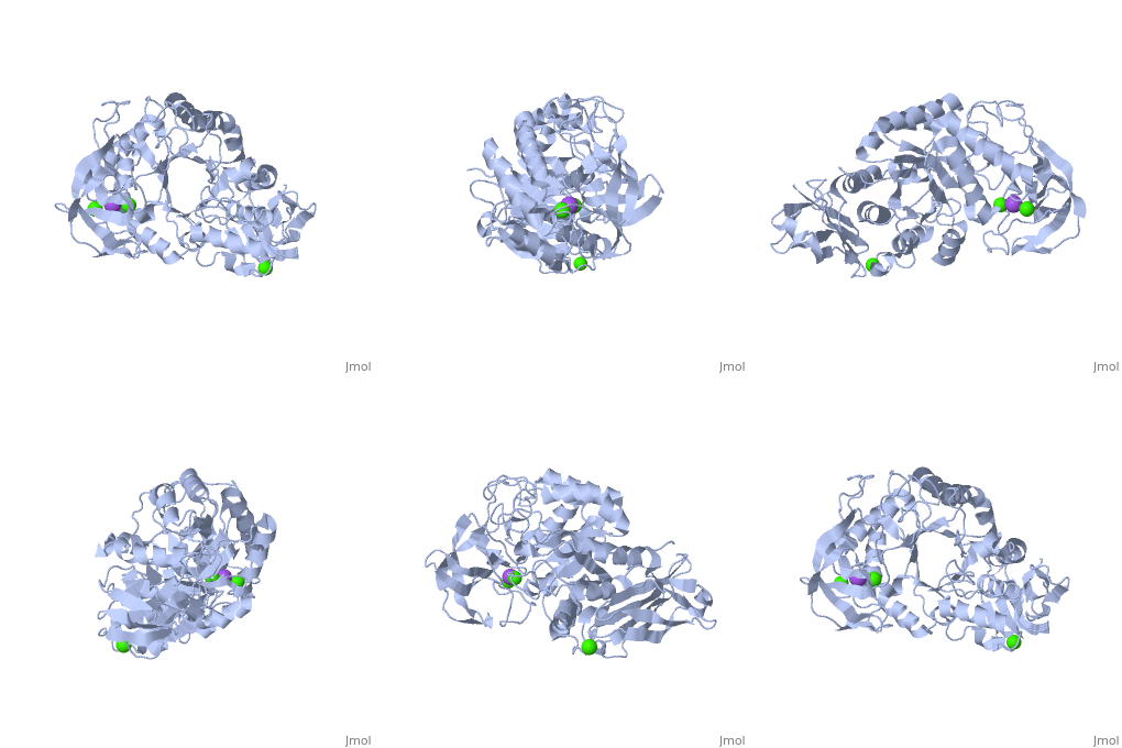
This is a default text for your page Alpha-amylase. Click above on edit this page to modify. Be careful with the < and > signs. You may include any references to papers as in: the use of JSmol in Proteopedia [1] or to the article describing Jmol [2] to the rescue. IntroductionThe (EC 3.2.1.1 ) (CAS# 9014-71-5) (alternative names: 1,4-α-D-glucan glucanohydrolase; glycogenase) are calcium metalloenzymes, completely unable to function in the absence of calcium. By acting at random locations along the starch chain, α-amylase breaks down long-chain carbohydrates, ultimately yielding maltotriose and maltose from amylose, or maltose, glucose and "limit dextrin" from amylopectin. Because it can act anywhere on the substrate, α-amylase tends to be faster-acting than β-amylase. In animals, it is a major digestive enzyme, and its optimum pH is 6.7–7.0.[3] In human physiology, both the salivary and pancreatic amylases are α-amylases. The α-amylases form is also found in plants, fungi (ascomycetes and basidiomycetes) and bacteria (Bacillus) DiseaseRelevanceStructural highlights
This is a sample scene created with SAT to by Group, and another to make of the protein. You can make your own scenes on SAT starting from scratch or loading and editing one of these sample scenes.
|
| ||||||||||
ReferencesReferences
- ↑ Hanson, R. M., Prilusky, J., Renjian, Z., Nakane, T. and Sussman, J. L. (2013), JSmol and the Next-Generation Web-Based Representation of 3D Molecular Structure as Applied to Proteopedia. Isr. J. Chem., 53:207-216. doi:http://dx.doi.org/10.1002/ijch.201300024
- ↑ Herraez A. Biomolecules in the computer: Jmol to the rescue. Biochem Mol Biol Educ. 2006 Jul;34(4):255-61. doi: 10.1002/bmb.2006.494034042644. PMID:21638687 doi:10.1002/bmb.2006.494034042644
