4ljo: Difference between revisions
No edit summary |
No edit summary |
||
| Line 2: | Line 2: | ||
<StructureSection load='4ljo' size='340' side='right' caption='[[4ljo]], [[Resolution|resolution]] 1.56Å' scene=''> | <StructureSection load='4ljo' size='340' side='right' caption='[[4ljo]], [[Resolution|resolution]] 1.56Å' scene=''> | ||
== Structural highlights == | == Structural highlights == | ||
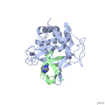
<table><tr><td colspan='2'>[[4ljo]] is a 2 chain structure with sequence from [http://en.wikipedia.org/wiki/ | <table><tr><td colspan='2'>[[4ljo]] is a 2 chain structure with sequence from [http://en.wikipedia.org/wiki/Bos_taurus Bos taurus] and [http://en.wikipedia.org/wiki/Human Human]. Full crystallographic information is available from [http://oca.weizmann.ac.il/oca-bin/ocashort?id=4LJO OCA]. For a <b>guided tour on the structure components</b> use [http://oca.weizmann.ac.il/oca-docs/fgij/fg.htm?mol=4LJO FirstGlance]. <br> | ||
</td></tr><tr id='ligand'><td class="sblockLbl"><b>[[Ligand|Ligands:]]</b></td><td class="sblockDat"><scene name='pdbligand=IMD:IMIDAZOLE'>IMD</scene>, <scene name='pdbligand=ZN:ZINC+ION'>ZN</scene></td></tr> | </td></tr><tr id='ligand'><td class="sblockLbl"><b>[[Ligand|Ligands:]]</b></td><td class="sblockDat"><scene name='pdbligand=IMD:IMIDAZOLE'>IMD</scene>, <scene name='pdbligand=ZN:ZINC+ION'>ZN</scene></td></tr> | ||
<tr id='related'><td class="sblockLbl"><b>[[Related_structure|Related:]]</b></td><td class="sblockDat">[[4ljp|4ljp]], [[4ljq|4ljq]]</td></tr> | <tr id='related'><td class="sblockLbl"><b>[[Related_structure|Related:]]</b></td><td class="sblockDat">[[4ljp|4ljp]], [[4ljq|4ljq]]</td></tr> | ||
| Line 23: | Line 23: | ||
__TOC__ | __TOC__ | ||
</StructureSection> | </StructureSection> | ||
[[Category: | [[Category: Bos taurus]] | ||
[[Category: Human]] | [[Category: Human]] | ||
[[Category: Brown, N R.]] | [[Category: Brown, N R.]] | ||
Revision as of 18:19, 12 October 2014
Structure of an active ligase (HOIP)/ubiquitin transfer complexStructure of an active ligase (HOIP)/ubiquitin transfer complex
Structural highlights
Publication Abstract from PubMedLinear ubiquitin chains are important regulators of cellular signalling pathways that control innate immunity and inflammation through nuclear factor (NF)-kappaB activation and protection against tumour necrosis factor-alpha-induced apoptosis. They are synthesized by HOIP, which belongs to the RBR (RING-between-RING) family of E3 ligases and is the catalytic component of LUBAC (linear ubiquitin chain assembly complex), a multisubunit E3 ligase. RBR family members act as RING/HECT hybrids, employing RING1 to recognize ubiquitin-loaded E2 while a conserved cysteine in RING2 subsequently forms a thioester intermediate with the transferred or 'donor' ubiquitin. Here we report the crystal structure of the catalytic core of HOIP in its apo form and in complex with ubiquitin. The carboxy-terminal portion of HOIP adopts a novel fold that, together with a zinc-finger, forms a ubiquitin-binding platform that orients the acceptor ubiquitin and positions its alpha-amino group for nucleophilic attack on the E3 approximately ubiquitin thioester. The C-terminal tail of a second ubiquitin molecule is located in close proximity to the catalytic cysteine, providing a unique snapshot of the ubiquitin transfer complex containing both donor and acceptor ubiquitin. These interactions are required for activation of the NF-kappaB pathway in vivo, and they explain the determinants of linear ubiquitin chain specificity by LUBAC. Structural basis for ligase-specific conjugation of linear ubiquitin chains by HOIP.,Stieglitz B, Rana RR, Koliopoulos MG, Morris-Davies AC, Schaeffer V, Christodoulou E, Howell S, Brown NR, Dikic I, Rittinger K Nature. 2013 Oct 20. doi: 10.1038/nature12638. PMID:24141947[1] From MEDLINE®/PubMed®, a database of the U.S. National Library of Medicine. See AlsoReferences
|
| ||||||||||||||||||||