1eyg: Difference between revisions
No edit summary |
No edit summary |
||
| Line 1: | Line 1: | ||
[[Image:1eyg.gif|left|200px]] | [[Image:1eyg.gif|left|200px]] | ||
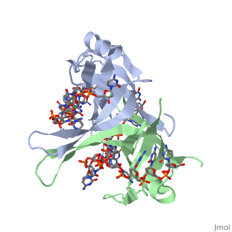
'''Crystal structure of chymotryptic fragment of E. coli ssb bound to two 35-mer single strand DNAS''' | {{Structure | ||
|PDB= 1eyg |SIZE=350|CAPTION= <scene name='initialview01'>1eyg</scene>, resolution 2.80Å | |||
|SITE= | |||
|LIGAND= | |||
|ACTIVITY= | |||
|GENE= | |||
}} | |||
'''Crystal structure of chymotryptic fragment of E. coli ssb bound to two 35-mer single strand DNAS''' | |||
==Overview== | ==Overview== | ||
| Line 7: | Line 16: | ||
==About this Structure== | ==About this Structure== | ||
1EYG is a [ | 1EYG is a [[Single protein]] structure of sequence from [http://en.wikipedia.org/wiki/Escherichia_coli Escherichia coli]. Full crystallographic information is available from [http://oca.weizmann.ac.il/oca-bin/ocashort?id=1EYG OCA]. | ||
==Reference== | ==Reference== | ||
Structure of the DNA binding domain of E. coli SSB bound to ssDNA., Raghunathan S, Kozlov AG, Lohman TM, Waksman G, Nat Struct Biol. 2000 Aug;7(8):648-52. PMID:[http:// | Structure of the DNA binding domain of E. coli SSB bound to ssDNA., Raghunathan S, Kozlov AG, Lohman TM, Waksman G, Nat Struct Biol. 2000 Aug;7(8):648-52. PMID:[http://www.ncbi.nlm.nih.gov/pubmed/10932248 10932248] | ||
[[Category: Escherichia coli]] | [[Category: Escherichia coli]] | ||
[[Category: Single protein]] | [[Category: Single protein]] | ||
| Line 17: | Line 26: | ||
[[Category: protein-dna complex; ob fold; se-met; mad phasing; ssb; binding mode]] | [[Category: protein-dna complex; ob fold; se-met; mad phasing; ssb; binding mode]] | ||
''Page seeded by [http://oca.weizmann.ac.il/oca OCA ] on Thu | ''Page seeded by [http://oca.weizmann.ac.il/oca OCA ] on Thu Mar 20 11:01:55 2008'' | ||
Revision as of 12:01, 20 March 2008
| |||||||
| , resolution 2.80Å | |||||||
|---|---|---|---|---|---|---|---|
| Coordinates: | save as pdb, mmCIF, xml | ||||||
Crystal structure of chymotryptic fragment of E. coli ssb bound to two 35-mer single strand DNAS
OverviewOverview
The structure of the homotetrameric DNA binding domain of the single stranded DNA binding protein from Escherichia coli (Eco SSB) bound to two 35-mer single stranded DNAs was determined to a resolution of 2.8 A. This structure describes the vast network of interactions that results in the extensive wrapping of single stranded DNA around the SSB tetramer and suggests a structural basis for its various binding modes.
About this StructureAbout this Structure
1EYG is a Single protein structure of sequence from Escherichia coli. Full crystallographic information is available from OCA.
ReferenceReference
Structure of the DNA binding domain of E. coli SSB bound to ssDNA., Raghunathan S, Kozlov AG, Lohman TM, Waksman G, Nat Struct Biol. 2000 Aug;7(8):648-52. PMID:10932248
Page seeded by OCA on Thu Mar 20 11:01:55 2008