1a9u: Difference between revisions
No edit summary |
No edit summary |
||
| Line 1: | Line 1: | ||
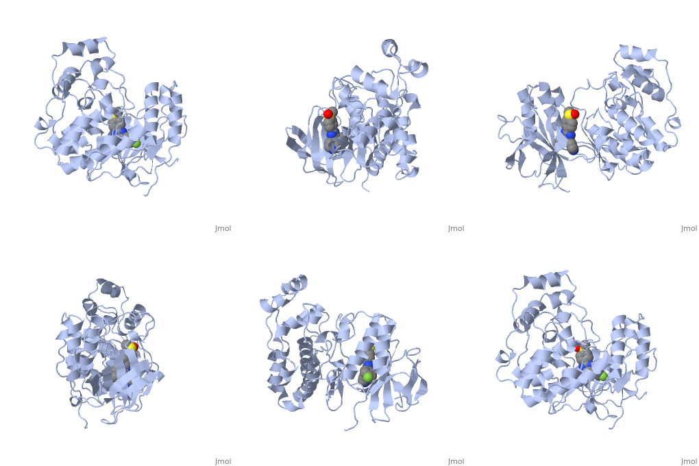
[[Image: | ==THE COMPLEX STRUCTURE OF THE MAP KINASE P38/SB203580== | ||
<StructureSection load='1a9u' size='340' side='right' caption='[[1a9u]], [[Resolution|resolution]] 2.50Å' scene=''> | |||
== Structural highlights == | |||
<table><tr><td colspan='2'>[[1a9u]] is a 1 chain structure with sequence from [http://en.wikipedia.org/wiki/Homo_sapiens Homo sapiens]. Full crystallographic information is available from [http://oca.weizmann.ac.il/oca-bin/ocashort?id=1A9U OCA]. For a <b>guided tour on the structure components</b> use [http://oca.weizmann.ac.il/oca-docs/fgij/fg.htm?mol=1A9U FirstGlance]. <br> | |||
</td></tr><tr><td class="sblockLbl"><b>[[Ligand|Ligands:]]</b></td><td class="sblockDat"><scene name='pdbligand=SB2:4-[5-(4-FLUORO-PHENYL)-2-(4-METHANESULFINYL-PHENYL)-3H-IMIDAZOL-4-YL]-PYRIDINE'>SB2</scene><br> | |||
<tr><td class="sblockLbl"><b>Resources:</b></td><td class="sblockDat"><span class='plainlinks'>[http://oca.weizmann.ac.il/oca-docs/fgij/fg.htm?mol=1a9u FirstGlance], [http://oca.weizmann.ac.il/oca-bin/ocaids?id=1a9u OCA], [http://www.rcsb.org/pdb/explore.do?structureId=1a9u RCSB], [http://www.ebi.ac.uk/pdbsum/1a9u PDBsum]</span></td></tr> | |||
<table> | |||
== Evolutionary Conservation == | |||
[[Image:Consurf_key_small.gif|200px|right]] | |||
Check<jmol> | |||
<jmolCheckbox> | |||
<scriptWhenChecked>select protein; define ~consurf_to_do selected; consurf_initial_scene = true; script "/wiki/ConSurf/a9/1a9u_consurf.spt"</scriptWhenChecked> | |||
<scriptWhenUnchecked>script /wiki/extensions/Proteopedia/spt/initialview01.spt</scriptWhenUnchecked> | |||
<text>to colour the structure by Evolutionary Conservation</text> | |||
</jmolCheckbox> | |||
</jmol>, as determined by [http://consurfdb.tau.ac.il/ ConSurfDB]. You may read the [[Conservation%2C_Evolutionary|explanation]] of the method and the full data available from [http://bental.tau.ac.il/new_ConSurfDB/chain_selection.php?pdb_ID=2ata ConSurf]. | |||
<div style="clear:both"></div> | |||
<div style="background-color:#fffaf0;"> | |||
== Publication Abstract from PubMed == | |||
BACKGROUND: The mitogen-activated protein (MAP) kinases are important signaling molecules that participate in diverse cellular events and are potential targets for intervention in inflammation, cancer, and other diseases. The MAP kinase p38 is responsive to environmental stresses and is involved in the production of cytokines during inflammation. In contrast, the activation of the MAP kinase ERK2 (extracellular-signal-regulated kinase 2) leads to cellular differentiation or proliferation. The anti-inflammatory agent pyridinylimidazole and its analogs (SB [SmithKline Beecham] compounds) are highly potent and selective inhibitors of p38, but not of the closely-related ERK2, or other serine/threonine kinases. Although these compounds are known to bind to the ATP-binding site, the origin of the inhibitory specificity toward p38 is not clear. RESULTS: We report the structural basis for the exceptional selectivity of these SB compounds for p38 over ERK2, as determined by comparative crystallography. In addition, structural data on the origin of olomoucine (a better inhibitor of ERK2) selectivity are presented. The crystal structures of four SB compounds in complex with p38 and of one SB compound and olomoucine in complex with ERK2 are presented here. The SB inhibitors bind in an extended pocket in the active site and are complementary to the open domain structure of the low-activity form of p38. The relatively closed domain structure of ERK2 is able to accommodate the smaller olomoucine. CONCLUSIONS: The unique kinase-inhibitor interactions observed in these complexes originate from amino-acid replacements in the active site and replacements distant from the active site that affect the size of the domain interface. This structural information should facilitate the design of better MAP-kinase inhibitors for the treatment of inflammation and other diseases. | |||
Structural basis of inhibitor selectivity in MAP kinases.,Wang Z, Canagarajah BJ, Boehm JC, Kassisa S, Cobb MH, Young PR, Abdel-Meguid S, Adams JL, Goldsmith EJ Structure. 1998 Sep 15;6(9):1117-28. PMID:9753691<ref>PMID:9753691</ref> | |||
From MEDLINE®/PubMed®, a database of the U.S. National Library of Medicine.<br> | |||
</div> | |||
==See Also== | ==See Also== | ||
*[[Mitogen-activated protein kinase|Mitogen-activated protein kinase]] | *[[Mitogen-activated protein kinase|Mitogen-activated protein kinase]] | ||
== References == | |||
== | <references/> | ||
< | __TOC__ | ||
</StructureSection> | |||
[[Category: Homo sapiens]] | [[Category: Homo sapiens]] | ||
[[Category: Abdel-Meguid, S.]] | [[Category: Abdel-Meguid, S.]] | ||
Revision as of 11:32, 23 July 2014
THE COMPLEX STRUCTURE OF THE MAP KINASE P38/SB203580THE COMPLEX STRUCTURE OF THE MAP KINASE P38/SB203580
Structural highlights
Evolutionary Conservation Check, as determined by ConSurfDB. You may read the explanation of the method and the full data available from ConSurf. Publication Abstract from PubMedBACKGROUND: The mitogen-activated protein (MAP) kinases are important signaling molecules that participate in diverse cellular events and are potential targets for intervention in inflammation, cancer, and other diseases. The MAP kinase p38 is responsive to environmental stresses and is involved in the production of cytokines during inflammation. In contrast, the activation of the MAP kinase ERK2 (extracellular-signal-regulated kinase 2) leads to cellular differentiation or proliferation. The anti-inflammatory agent pyridinylimidazole and its analogs (SB [SmithKline Beecham] compounds) are highly potent and selective inhibitors of p38, but not of the closely-related ERK2, or other serine/threonine kinases. Although these compounds are known to bind to the ATP-binding site, the origin of the inhibitory specificity toward p38 is not clear. RESULTS: We report the structural basis for the exceptional selectivity of these SB compounds for p38 over ERK2, as determined by comparative crystallography. In addition, structural data on the origin of olomoucine (a better inhibitor of ERK2) selectivity are presented. The crystal structures of four SB compounds in complex with p38 and of one SB compound and olomoucine in complex with ERK2 are presented here. The SB inhibitors bind in an extended pocket in the active site and are complementary to the open domain structure of the low-activity form of p38. The relatively closed domain structure of ERK2 is able to accommodate the smaller olomoucine. CONCLUSIONS: The unique kinase-inhibitor interactions observed in these complexes originate from amino-acid replacements in the active site and replacements distant from the active site that affect the size of the domain interface. This structural information should facilitate the design of better MAP-kinase inhibitors for the treatment of inflammation and other diseases. Structural basis of inhibitor selectivity in MAP kinases.,Wang Z, Canagarajah BJ, Boehm JC, Kassisa S, Cobb MH, Young PR, Abdel-Meguid S, Adams JL, Goldsmith EJ Structure. 1998 Sep 15;6(9):1117-28. PMID:9753691[1] From MEDLINE®/PubMed®, a database of the U.S. National Library of Medicine. See AlsoReferences |
| ||||||||||||||||
