2ren: Difference between revisions
No edit summary |
No edit summary |
||
| Line 4: | Line 4: | ||
==Overview== | ==Overview== | ||
The x-ray crystal structure of recombinant human renin has been | The x-ray crystal structure of recombinant human renin has been determined. Molecular dynamics techniques that included crystallographic data as a restraint were used to improve an initial model based on porcine pepsinogen. The present agreement factor for data from 8.0 to 2.5 angstroms (A) is 0.236. Some of the surface loops are poorly determined, and these disordered regions border a 30 A wide solvent channel. Comparison of renin with other aspartyl proteinases shows that, although the structural cores and active sites are highly conserved, surface residues, some of which are critical for specificity, vary greatly (up to 10A). Knowledge of the actual structure, as opposed to the use of models based on related enzymes, should facilitate the design of renin inhibitors. | ||
==Disease== | ==Disease== | ||
| Line 17: | Line 17: | ||
[[Category: Renin]] | [[Category: Renin]] | ||
[[Category: Single protein]] | [[Category: Single protein]] | ||
[[Category: James, M | [[Category: James, M N.G.]] | ||
[[Category: Sielecki, A | [[Category: Sielecki, A R.]] | ||
[[Category: NAG]] | [[Category: NAG]] | ||
[[Category: hydrolase(acid proteinase)]] | [[Category: hydrolase(acid proteinase)]] | ||
''Page seeded by [http://oca.weizmann.ac.il/oca OCA ] on | ''Page seeded by [http://oca.weizmann.ac.il/oca OCA ] on Thu Feb 21 18:46:42 2008'' | ||
Revision as of 19:46, 21 February 2008
|
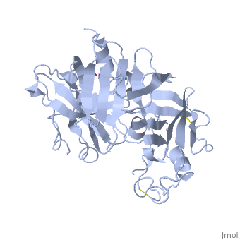
STRUCTURE OF RECOMBINANT HUMAN RENIN, A TARGET FOR CARDIOVASCULAR-ACTIVE DRUGS, AT 2.5 ANGSTROMS RESOLUTION
OverviewOverview
The x-ray crystal structure of recombinant human renin has been determined. Molecular dynamics techniques that included crystallographic data as a restraint were used to improve an initial model based on porcine pepsinogen. The present agreement factor for data from 8.0 to 2.5 angstroms (A) is 0.236. Some of the surface loops are poorly determined, and these disordered regions border a 30 A wide solvent channel. Comparison of renin with other aspartyl proteinases shows that, although the structural cores and active sites are highly conserved, surface residues, some of which are critical for specificity, vary greatly (up to 10A). Knowledge of the actual structure, as opposed to the use of models based on related enzymes, should facilitate the design of renin inhibitors.
DiseaseDisease
Known diseases associated with this structure: Hyperproreninemia OMIM:[179820], Renal tubular dysgenesis OMIM:[179820]
About this StructureAbout this Structure
2REN is a Single protein structure of sequence from Homo sapiens with as ligand. Active as Renin, with EC number 3.4.23.15 Full crystallographic information is available from OCA.
ReferenceReference
Structure of recombinant human renin, a target for cardiovascular-active drugs, at 2.5 A resolution., Sielecki AR, Hayakawa K, Fujinaga M, Murphy ME, Fraser M, Muir AK, Carilli CT, Lewicki JA, Baxter JD, James MN, Science. 1989 Mar 10;243(4896):1346-51. PMID:2493678
Page seeded by OCA on Thu Feb 21 18:46:42 2008