1ihr: Difference between revisions
New page: left|200px<br /><applet load="1ihr" size="450" color="white" frame="true" align="right" spinBox="true" caption="1ihr, resolution 1.55Å" /> '''Crystal structure of... |
No edit summary |
||
| Line 1: | Line 1: | ||
[[Image:1ihr.gif|left|200px]]<br /><applet load="1ihr" size=" | [[Image:1ihr.gif|left|200px]]<br /><applet load="1ihr" size="350" color="white" frame="true" align="right" spinBox="true" | ||
caption="1ihr, resolution 1.55Å" /> | caption="1ihr, resolution 1.55Å" /> | ||
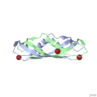
'''Crystal structure of the dimeric C-terminal domain of TonB'''<br /> | '''Crystal structure of the dimeric C-terminal domain of TonB'''<br /> | ||
==Overview== | ==Overview== | ||
The TonB-dependent complex of Gram-negative bacteria couples the inner | The TonB-dependent complex of Gram-negative bacteria couples the inner membrane proton motive force to the active transport of iron.siderophore and vitamin B(12) across the outer membrane. The structural basis of that process has not been described so far in full detail. The crystal structure of the C-terminal domain of TonB from Escherichia coli has now been solved by multiwavelength anomalous diffraction and refined at 1.55-A resolution, providing the first evidence that this region of TonB (residues 164-239) dimerizes. Moreover, the structure shows a novel architecture that has no structural homologs among any known proteins. The dimer of the C-terminal domain of TonB is cylinder-shaped with a length of 65 A and a diameter of 25 A. Each monomer contains three beta strands and a single alpha helix. The two monomers are intertwined with each other, and all six beta-strands of the dimer make a large antiparallel beta-sheet. We propose a plausible model of binding of TonB to FhuA and FepA, two TonB-dependent outer-membrane receptors. | ||
==About this Structure== | ==About this Structure== | ||
1IHR is a [http://en.wikipedia.org/wiki/Single_protein Single protein] structure of sequence from [http://en.wikipedia.org/wiki/Escherichia_coli Escherichia coli] with BR as [http://en.wikipedia.org/wiki/ligand ligand]. Full crystallographic information is available from [http:// | 1IHR is a [http://en.wikipedia.org/wiki/Single_protein Single protein] structure of sequence from [http://en.wikipedia.org/wiki/Escherichia_coli Escherichia coli] with <scene name='pdbligand=BR:'>BR</scene> as [http://en.wikipedia.org/wiki/ligand ligand]. Full crystallographic information is available from [http://oca.weizmann.ac.il/oca-bin/ocashort?id=1IHR OCA]. | ||
==Reference== | ==Reference== | ||
| Line 21: | Line 21: | ||
[[Category: novel fold]] | [[Category: novel fold]] | ||
''Page seeded by [http:// | ''Page seeded by [http://oca.weizmann.ac.il/oca OCA ] on Thu Feb 21 13:11:59 2008'' | ||
Revision as of 14:12, 21 February 2008
|
Crystal structure of the dimeric C-terminal domain of TonB
OverviewOverview
The TonB-dependent complex of Gram-negative bacteria couples the inner membrane proton motive force to the active transport of iron.siderophore and vitamin B(12) across the outer membrane. The structural basis of that process has not been described so far in full detail. The crystal structure of the C-terminal domain of TonB from Escherichia coli has now been solved by multiwavelength anomalous diffraction and refined at 1.55-A resolution, providing the first evidence that this region of TonB (residues 164-239) dimerizes. Moreover, the structure shows a novel architecture that has no structural homologs among any known proteins. The dimer of the C-terminal domain of TonB is cylinder-shaped with a length of 65 A and a diameter of 25 A. Each monomer contains three beta strands and a single alpha helix. The two monomers are intertwined with each other, and all six beta-strands of the dimer make a large antiparallel beta-sheet. We propose a plausible model of binding of TonB to FhuA and FepA, two TonB-dependent outer-membrane receptors.
About this StructureAbout this Structure
1IHR is a Single protein structure of sequence from Escherichia coli with as ligand. Full crystallographic information is available from OCA.
ReferenceReference
Crystal structure of the dimeric C-terminal domain of TonB reveals a novel fold., Chang C, Mooser A, Pluckthun A, Wlodawer A, J Biol Chem. 2001 Jul 20;276(29):27535-40. Epub 2001 Apr 27. PMID:11328822
Page seeded by OCA on Thu Feb 21 13:11:59 2008