P53-DNA Recognition: Difference between revisions
No edit summary |
No edit summary |
||
| Line 8: | Line 8: | ||
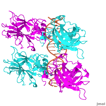
[[Image:p53-intro.jpg|thumb|left|300px|Figure 1: Crystal structure of a p53 DBD tetramer-DNA complex; PDB ID# 3KZ8 <ref name='kitayner'>Kitayner M, Rozenberg H, Rohs R, Suad O, Rabinovich D, Honig B, Shakked Z. Diversity in DNA recognition by p53 revealed by crystal structures with Hoogsteen base pairs. Nat Struct Mol Biol. 2010;17(4):423-9.</ref>.]] | [[Image:p53-intro.jpg|thumb|left|300px|Figure 1: Crystal structure of a p53 DBD tetramer-DNA complex; PDB ID# 3KZ8 <ref name='kitayner'>Kitayner M, Rozenberg H, Rohs R, Suad O, Rabinovich D, Honig B, Shakked Z. Diversity in DNA recognition by p53 revealed by crystal structures with Hoogsteen base pairs. Nat Struct Mol Biol. 2010;17(4):423-9.</ref>.]] | ||
[[Image:p53-consensus.jpg|thumb|right|400px|consensus site]] | |||
[[Image:p53-domains.jpg|thumb|right|300px|domains]] | [[Image:p53-domains.jpg|thumb|right|300px|domains]] | ||
Also known as the '''Guardian of the Genome''', p53 is | Also known as the '''Guardian of the Genome''', the tumor suppressor p53 is central in the natural defense against human cancer. The protein is activated by stress factors that can compromise the genomic integrity of the cell, and this activation unleashes the function of p53 as transcription factor. It binds as a tetramer (Figure 1) to a large range of DNA response elements, which are formed by two decameric half-sites that are separated by a variable number of base pairs (Figure2). Binding of p53 to different response elements leads to distinct biological responses, such as cell-cycle arrest, senescence or apoptosis. | ||
Revision as of 23:00, 3 July 2012
| This Sandbox is Reserved from 22 April 2012, through 31 August 2012 for use in the course "Protein DNA" taught by Remo_Rohs at the La Canada High School, USA. This reservation includes Sandbox Reserved 169 through Sandbox Reserved 170. |
To get started:
More help: Help:Editing |
A Base Pairing Variant Enhances p53 Binding to a Response ElementA Base Pairing Variant Enhances p53 Binding to a Response Element
Introduction and Biological Role of the Tumor Suppressor p53Introduction and Biological Role of the Tumor Suppressor p53


Also known as the Guardian of the Genome, the tumor suppressor p53 is central in the natural defense against human cancer. The protein is activated by stress factors that can compromise the genomic integrity of the cell, and this activation unleashes the function of p53 as transcription factor. It binds as a tetramer (Figure 1) to a large range of DNA response elements, which are formed by two decameric half-sites that are separated by a variable number of base pairs (Figure2). Binding of p53 to different response elements leads to distinct biological responses, such as cell-cycle arrest, senescence or apoptosis.
|


Further ReadingFurther Reading
A more general discussion of structural origins of binding specificity in protein-DNA recognition has been published along with a suggestion for a new classification of protein-DNA readout modes that goes beyond the historical description of direct and indirect readout[2].
AcknowledgementsAcknowledgements
This Proteopedia page originates from the partnership of the Rohs Laboratory at the University of Southern California with La Canada High School. This partnership was initiated by Remo Rohs and Patty Compeau in September 2011 as Bioinformatics Institute, which is part of the Institutes of the 21st Century. Contributors to this page are USC graduate students Ana Carolina Dantas Machado, Proteopedia editor Eran Hodis, and La Canada High School students xxx. Research presented in this article has been conducted in the Shakked Lab at the Weizmann Institute of Science, the Rohs and L. Chen Labs at USC, and the Honig Lab at Columbia University. Furthermore, technical help by Proteopedia editors Eran Hodis, Eric Martz, Jaime Prilusky, and Joel Sussman is acknowledged.
ReferencesReferences
- ↑ Kitayner M, Rozenberg H, Rohs R, Suad O, Rabinovich D, Honig B, Shakked Z. Diversity in DNA recognition by p53 revealed by crystal structures with Hoogsteen base pairs. Nat Struct Mol Biol. 2010;17(4):423-9.
- ↑ Rohs R, Jin X, West SM, Joshi R, Honig B, Mann RS. Origins of specificity in protein-DNA recognition. Annu Rev Biochem. 2010;79:233-69.