Avidin: Difference between revisions
Michal Harel (talk | contribs) No edit summary |
m Strepavidin - Streptavidin |
||
| Line 3: | Line 3: | ||
'''3D Structure of Avidin''' | '''3D Structure of Avidin''' | ||
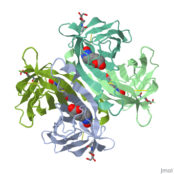
[[Avidin]] '''(Av)''' is a protein found in egg white which binds biotin (vitamin B7) with high affinity. This property makes it a powerful tool in various protein pull-down assays. Similar proteins are | [[Avidin]] '''(Av)''' is a protein found in egg white which binds biotin (vitamin B7) with high affinity. This property makes it a powerful tool in various protein pull-down assays. Similar proteins are streptavidin (StrAv) from the bacterium ''Streptomyces avidinii'', Rhizavidin (RhiAv) from ''Rhizobium etli'', Xenavidin from frog, and the avidin-related proteins AVR2 and AVR4. The N-acetylglucosamine sugars are shown as space filling objects in the image on the left. | ||
Avidin is one of the [[CBI Molecules]] being studied in the [http://www.umass.edu/cbi/ University of Massachusetts Amherst Chemistry-Biology Interface Program] at UMass Amherst and on display at the [http://www.molecularplayground.org/ Molecular Playground]. | Avidin is one of the [[CBI Molecules]] being studied in the [http://www.umass.edu/cbi/ University of Massachusetts Amherst Chemistry-Biology Interface Program] at UMass Amherst and on display at the [http://www.molecularplayground.org/ Molecular Playground]. | ||
| Line 37: | Line 37: | ||
== | == Streptavidin == | ||
| Line 45: | Line 45: | ||
[[1swh]], [[1swj]], [[1swl]], [[1swo]], [[1swq]], [[1sws]], [[2y3e]] - StrAv core (mutant)<br /> | [[1swh]], [[1swj]], [[1swl]], [[1swo]], [[1swq]], [[1sws]], [[2y3e]] - StrAv core (mutant)<br /> | ||
== | == Streptavidin binary complexes== | ||
[[1rxh]] – StrAv (mutant)+ biotinyl p-nitroaniline <br /> | [[1rxh]] – StrAv (mutant)+ biotinyl p-nitroaniline <br /> | ||
Revision as of 14:38, 14 November 2011
Template:STRUCTURE 1ave 3D Structure of Avidin
Avidin (Av) is a protein found in egg white which binds biotin (vitamin B7) with high affinity. This property makes it a powerful tool in various protein pull-down assays. Similar proteins are streptavidin (StrAv) from the bacterium Streptomyces avidinii, Rhizavidin (RhiAv) from Rhizobium etli, Xenavidin from frog, and the avidin-related proteins AVR2 and AVR4. The N-acetylglucosamine sugars are shown as space filling objects in the image on the left.
Avidin is one of the CBI Molecules being studied in the University of Massachusetts Amherst Chemistry-Biology Interface Program at UMass Amherst and on display at the Molecular Playground.
Avidin is a tetrameric protein what in different words means that if we could, we would see a protein with four regions that look identical one to the other. However, to isolate the four regions as a whole in order to show the protein three dimensional structure has been quite diffucult so far and we just can see the three dimensional structure for two of those four units. [xtra 1][xtra 2]
The binding affinity of biotin for the avidin is very hight or in other words, the strength of the interations between the biotin and the avidin receptors is so strong that washing the biotin-avidin complex is not enough to remove the ligand from its pocket. This is not even possible by adding more biotin molecules to the system avidin-biotin. Once a biotin has bound a pocket in the avidin, it is almost imposible to remove it in a biologica system!
|
Each monomer is an eight-stranded antiparallel . Binding of biotin involves a highly stabilized network of polar and hydrophobic interactions.
The presence of additional hydrophobic and hydrophilic groups in the binding site of avidin may account for its higher affinity constant.
Unexpectedly, a residual N-acetylglucosamine moiety was detected in the deglycosylated avidin monomer. These appear along with the biotin but outside of the biotin binding pockets.
About this StructureAbout this Structure
2AVI is a 2 chains structure of sequences from Gallus gallus. Full crystallographic information is available from OCA.
3D structures of Avidin3D structures of Avidin
Update November 2011
3fdc, 2jgs, 2c4i, 1vyo, 1rav, 1ave – cAv – chicken
2a5b, 2a5c, 2a8g – cAv+deoxyribose
1nqn, 2cam – cAv (mutant)
1ldo, 1ldq, 1lel, 1ij8, 1avd, 2avi – cAV + biotin derivative
StreptavidinStreptavidin
2bc3, 2iza, 2izb, 2izc, 2izd, 2ize, 2rta, 2rtb, 2rtc - StrAv - Streptomyces avidinii
3mm0, 3pk2, 3rds, 3rdx, 3re5, 3re6 - StrAv (mutant)
1swa, 1swb, 1swc, 1ry1 - StrAv core
1swh, 1swj, 1swl, 1swo, 1swq, 1sws, 2y3e - StrAv core (mutant)
Streptavidin binary complexesStreptavidin binary complexes
1rxh – StrAv (mutant)+ biotinyl p-nitroaniline
1rxj, 1rxk, 1nqm, 1kff, 1kl4, 1luq, 1mm9, 1moy, 1n4j, 1n7y, 1n9y, 1nbx, 1swf, 1swu,
1kl3, 1kl5 - StrAv (mutant) + strep-tag II peptide
1rst, 1rsu - StrAv + strep-tag II peptide
1df8, 1mep, 1n43, 1n9m, 1ndj, 1swg, 3rdm, 3rdo - StrAv (mutant)+biotin
1nc9, 3rdq - StrAv (mutant)+biotin derivative
2izl, 2rtl, 2rtm, 2rtn, 2rto, 2rtp, 2rtq, 2rtr - StrAv +iminobiotin
1mk5, 1swd, 1swe, 2f01, 2gh7, 3ry2 - StrAv core+biotin
1swk, 1swn, 1swp, 1swr, 1swt, 3mg5, 2y3f - StrAv core (mutant) + biotin
1stp, 2izf, 2izg, 2izh, 2izi, 2izj, 2rtd, 2rte, 2rtf, 2rtg - StrAv +biotin
3rdu - StrAv (mutant) + PEG
1hqq, 1hxl – StrAv + MP-2 (mutant)
1hy2 - StrAv + MP-1
1hxz - StrAv + MP-2
1pts, 1sld, 1sle, 1slg, 1str, 1sts, 1vwa, 1vwb, 1vwc, 1vwd, 1vwe, 1vwf, 1vwg, 1vwh, 1vwi, 1vwj, 1vwk, 1vwl, 1vwm, 1vwn, 1vwo, 1vwp, 1vwq, 1vwr, 2g5l - StrAv + peptide
1slf - StrAv + SO4
1sre, 1srf, 1srg, 1srh, 1sri, 1srj - StrAv + benzoic acid ligands
1lcv, 1lcw, 1lcz, 1i9h – StrAv+biotin derivative
2izk, 2rth, 2rti, 2rtj, 2rtk – StrAV+glycouril
2qcb, 2wpu – StrAv+Ru ligand
RhizavidinRhizavidin
3ew1 – RhiAv – Rhizobium etli
3ew2 – RhiAV+biotin
TamavidinTamavidin
2zsc – TanAv – Tamogitake mushroom
AVR2AVR2
1wbi – cAVR2
AVR4AVR4
2fhl – cAVR4+biotinyl p-nitroaniline
2of9, 2ofa, 1y53 - cAVR4 (mutant)
2fhn, 2of8 - cAVR4 (mutant)+biotinyl p-nitroaniline
1y52, 1y55 - cAVR4 (mutant)+biotin
ReferenceReference
- ↑ Livnah O, Bayer EA, Wilchek M, Sussman JL. Three-dimensional structures of avidin and the avidin-biotin complex. Proc Natl Acad Sci U S A. 1993 Jun 1;90(11):5076-80. PMID:8506353
- ↑ Livnah O, Bayer EA, Wilchek M, Sussman JL. The structure of the complex between avidin and the dye, 2-(4'-hydroxyazobenzene) benzoic acid (HABA). FEBS Lett. 1993 Aug 9;328(1-2):165-8. PMID:8344421