2vgb: Difference between revisions
New page: left|200px<br /><applet load="2vgb" size="450" color="white" frame="true" align="right" spinBox="true" caption="2vgb, resolution 2.73Å" /> '''HUMAN ERYTHROCYTE PY... |
No edit summary |
||
| Line 1: | Line 1: | ||
[[Image:2vgb.jpg|left|200px]]<br /><applet load="2vgb" size=" | [[Image:2vgb.jpg|left|200px]]<br /><applet load="2vgb" size="350" color="white" frame="true" align="right" spinBox="true" | ||
caption="2vgb, resolution 2.73Å" /> | caption="2vgb, resolution 2.73Å" /> | ||
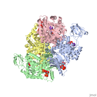
'''HUMAN ERYTHROCYTE PYRUVATE KINASE'''<br /> | '''HUMAN ERYTHROCYTE PYRUVATE KINASE'''<br /> | ||
| Line 7: | Line 7: | ||
==About this Structure== | ==About this Structure== | ||
2VGB is a [http://en.wikipedia.org/wiki/Single_protein Single protein] structure of sequence from [http://en.wikipedia.org/wiki/Homo_sapiens Homo sapiens] with FBP, PGA, K and MN as [http://en.wikipedia.org/wiki/ligands ligands]. This structure superseeds the now removed PDB entry 1LIU. Active as [http://en.wikipedia.org/wiki/Pyruvate_kinase Pyruvate kinase], with EC number [http://www.brenda-enzymes.info/php/result_flat.php4?ecno=2.7.1.40 2.7.1.40] | 2VGB is a [http://en.wikipedia.org/wiki/Single_protein Single protein] structure of sequence from [http://en.wikipedia.org/wiki/Homo_sapiens Homo sapiens] with <scene name='pdbligand=FBP:'>FBP</scene>, <scene name='pdbligand=PGA:'>PGA</scene>, <scene name='pdbligand=K:'>K</scene> and <scene name='pdbligand=MN:'>MN</scene> as [http://en.wikipedia.org/wiki/ligands ligands]. This structure superseeds the now removed PDB entry 1LIU. Active as [http://en.wikipedia.org/wiki/Pyruvate_kinase Pyruvate kinase], with EC number [http://www.brenda-enzymes.info/php/result_flat.php4?ecno=2.7.1.40 2.7.1.40] Known structural/functional Sites: <scene name='pdbsite=AC1:Fbp Binding Site For Chain A'>AC1</scene>, <scene name='pdbsite=AC2:Pga Binding Site For Chain A'>AC2</scene>, <scene name='pdbsite=AC3:K Binding Site For Chain A'>AC3</scene>, <scene name='pdbsite=AC4:Mn Binding Site For Chain A'>AC4</scene>, <scene name='pdbsite=AC5:Fbp Binding Site For Chain B'>AC5</scene>, <scene name='pdbsite=AC6:Pga Binding Site For Chain B'>AC6</scene>, <scene name='pdbsite=AC7:K Binding Site For Chain B'>AC7</scene>, <scene name='pdbsite=AC8:Mn Binding Site For Chain B'>AC8</scene>, <scene name='pdbsite=AC9:Fbp Binding Site For Chain C'>AC9</scene>, <scene name='pdbsite=BC1:Pga Binding Site For Chain C'>BC1</scene>, <scene name='pdbsite=BC2:K Binding Site For Chain C'>BC2</scene>, <scene name='pdbsite=BC3:Mn Binding Site For Chain C'>BC3</scene>, <scene name='pdbsite=BC4:Fbp Binding Site For Chain D'>BC4</scene>, <scene name='pdbsite=BC5:Pga Binding Site For Chain D'>BC5</scene>, <scene name='pdbsite=BC6:K Binding Site For Chain D'>BC6</scene> and <scene name='pdbsite=BC7:Mn Binding Site For Chain D'>BC7</scene>. Full crystallographic information is available from [http://oca.weizmann.ac.il/oca-bin/ocashort?id=2VGB OCA]. | ||
==Reference== | ==Reference== | ||
| Line 40: | Line 40: | ||
[[Category: transferase]] | [[Category: transferase]] | ||
''Page seeded by [http:// | ''Page seeded by [http://oca.weizmann.ac.il/oca OCA ] on Wed Jan 23 12:11:12 2008'' | ||
Revision as of 13:11, 23 January 2008
|
HUMAN ERYTHROCYTE PYRUVATE KINASE
OverviewOverview
Deficiency of human erythrocyte isozyme (RPK) is, together with, glucose-6-phosphate dehydrogenase deficiency, the most common cause of the, nonspherocytic hemolytic anemia. To provide a molecular framework to the, disease, we have solved the 2.7 A resolution crystal structure of human, RPK in complex with fructose 1,6-bisphosphate, the allosteric activator, and phosphoglycolate, a substrate analogue, and we have functionally and, structurally characterized eight mutants (G332S, G364D, T384M, D390N, R479H, R486W, R504L, and R532W) found in RPK-deficient patients. The, mutations target distinct regions of RPK structure, including domain, interfaces and catalytic and allosteric sites. The mutations affect to a, different extent thermostability, catalytic efficiency, and regulatory, properties. These studies are the first to correlate the clinical symptoms, with the molecular properties of the mutant enzymes. Mutations greatly, impairing thermostability and/or activity are associated with severe, anemia. Some mutant proteins exhibit moderate changes in the kinetic, parameters, which are sufficient to cause mild to severe anemia, underlining the crucial role of RPK for erythrocyte metabolism. Prediction, of the effects of mutations is difficult because there is no relation, between the nature and location of the replaced amino acid and the type of, molecular perturbation. Characterization of mutant proteins may serve as a, valuable tool to assist with diagnosis and genetic counseling.
About this StructureAbout this Structure
2VGB is a Single protein structure of sequence from Homo sapiens with , , and as ligands. This structure superseeds the now removed PDB entry 1LIU. Active as Pyruvate kinase, with EC number 2.7.1.40 Known structural/functional Sites: , , , , , , , , , , , , , , and . Full crystallographic information is available from OCA.
ReferenceReference
Structure and function of human erythrocyte pyruvate kinase. Molecular basis of nonspherocytic hemolytic anemia., Valentini G, Chiarelli LR, Fortin R, Dolzan M, Galizzi A, Abraham DJ, Wang C, Bianchi P, Zanella A, Mattevi A, J Biol Chem. 2002 Jun 28;277(26):23807-14. Epub 2002 Apr 17. PMID:11960989
Page seeded by OCA on Wed Jan 23 12:11:12 2008
Proteopedia Page Contributors and Editors (what is this?)Proteopedia Page Contributors and Editors (what is this?)
OCA- Pages with broken file links
- Homo sapiens
- Pyruvate kinase
- Single protein
- Abraham, D.J.
- Bianchi, P.
- Chiarelli, L.
- Dolzan, M.
- Fortin, R.
- Galizzi, A.
- Mattevi, A.
- Valentini, G.
- Wang, C.
- Zanella, A.
- FBP
- K
- MN
- PGA
- Alternative splicing
- Disease mutation
- Glycolysis
- Kinase
- Magnesium
- Metal-binding
- Phosphorylation
- Polymorphism
- Pyruvate
- Pyruvate kinase in the active r-state
- Transferase