User:Wayne Decatur/3fpn Morph methods: Difference between revisions
mNo edit summary |
mNo edit summary |
||
| Line 96: | Line 96: | ||
<nowiki>[</nowiki>Control the animation with the 'animation' submenu on the menu that comes up if you click on the Jmol frank in the bottom rigth corner. Also, if the animation seems to be stuck, scroll in the bar on the right of your browser.<nowiki>]</nowiki> | <nowiki>[</nowiki>Control the animation with the 'animation' submenu on the menu that comes up if you click on the Jmol frank in the bottom rigth corner. Also, if the animation seems to be stuck, scroll in the bar on the right of your browser.<nowiki>]</nowiki> | ||
<scene name='User:Wayne_Decatur/3fpn_Morph_methods/Testmodelallnew/1'>Test with new 3fpn file</scene> | |||
<scene name='User:Wayne_Decatur/3fpn_Morph_methods/Testmodelallribo/1'>Test of model all with ribosome file</scene> | <scene name='User:Wayne_Decatur/3fpn_Morph_methods/Testmodelallribo/1'>Test of model all with ribosome file</scene> | ||
Revision as of 22:12, 16 February 2010
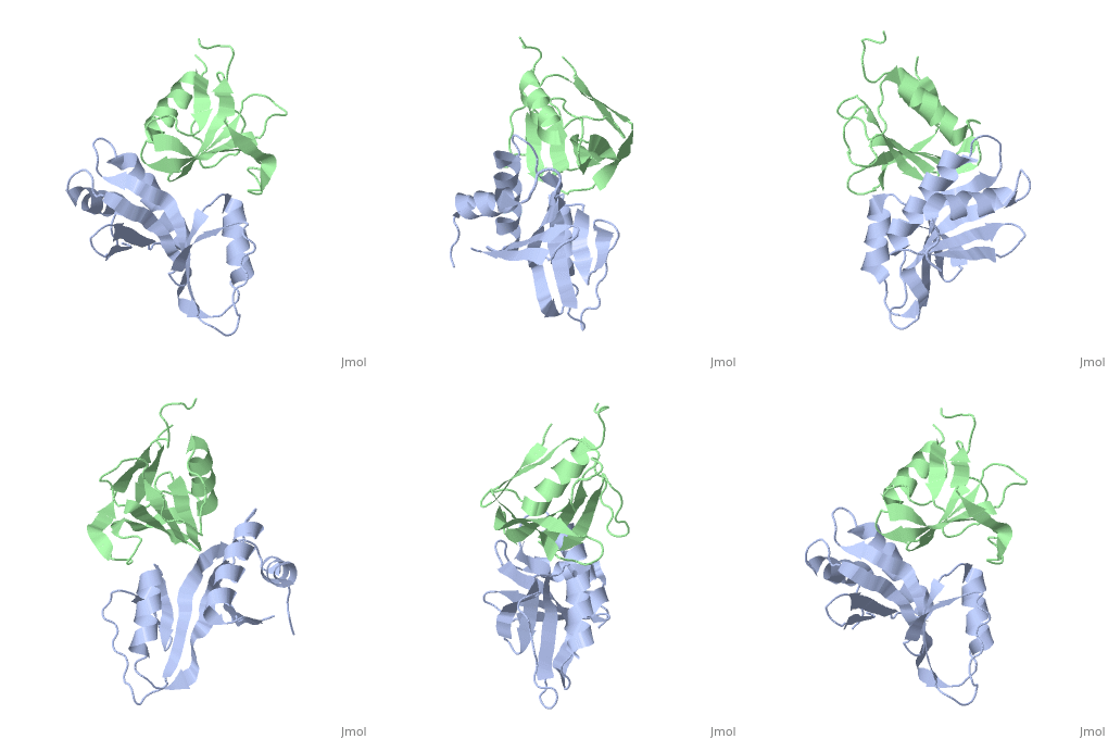
Moving to match Figure 3Moving to match Figure 3
Using Pymol and the 3fpn file, I moved so interface is perpendicular to y axis:
translate [10,0,0], chain b
rotate y, 65, chain b
Saved molecule.
Morph from normal 3fpn structure to view in Figure 3 of article describing the structureMorph from normal 3fpn structure to view in Figure 3 of article describing the structure
|
Took the two files and submitted them. Since the structures didn't have nucleic acids, I took the advice here and used the Yale Morph Server for morphing complexes. I got the e-mail and followed the link to a Jmol animation of the morph. I right clicked on the Jmol frank and click the top entry in the menu and then the bottom spot in the menu that came up to download the structures in the morph.
Uploaded to Proteopedia File:3fpntorotatedversion.pdb.
loaded '3fpntorotatedversion.pdb' in Scene Authoring Tools.
[Control the animation with the 'animation' submenu on the menu that comes up if you click on the Jmol frank in the bottom rigth corner. Also, if the animation seems to be stuck, scroll in the bar on the right of your browser.]
Paper on the structurePaper on the structure
- ↑ Pakotiprapha D, Liu Y, Verdine GL, Jeruzalmi D. A structural model for the damage-sensing complex in bacterial nucleotide excision repair. J Biol Chem. 2009 May 8;284(19):12837-44. Epub 2009 Mar 13. PMID:19287003 doi:10.1074/jbc.M900571200
