Rebecca Martin/Sandbox1: Difference between revisions
Jump to navigation
Jump to search
No edit summary |
No edit summary |
||
| Line 3: | Line 3: | ||
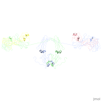
{{STRUCTURE_3cm9 | PDB=3cm9 | SCENE= IgA1}} | {{STRUCTURE_3cm9 | PDB=3cm9 | SCENE= IgA1}} | ||
| Line 15: | Line 26: | ||
== IgA1 and IgA2 == | == IgA1 and IgA2 == | ||
<applet load=' | <applet load='1r70' size='300' frame='true' align='right' caption='IgA2' /> <applet load='1iga' size='300' frame='true' align='right' caption='IgA1' /> | ||
Revision as of 16:52, 22 April 2009
IntroductionIntroduction
IgA1
General StructureGeneral Structure
- Antibody Structure
- Dimeric Structure
- Secretory Component
IgA1 and IgA2IgA1 and IgA2
|
|
Secretory Component and Polyimmunoglobulin receptorSecretory Component and Polyimmunoglobulin receptor
|
Insights into FunctionInsights into Function
EvolutionEvolution
Implications in Science and MedicineImplications in Science and Medicine
Limitations of the Current StudiesLimitations of the Current Studies
Questions for the FutureQuestions for the Future
LinksLinks
IgAIgA
- Model of human IgA1 determined by solution scattering, curve-fitting, and homology modeling 1iga
- Model of human IgA2 determined by solution scattering, curve fitting and homology modelling 1r70
- Crystal structure of human FcaRI bound to IgA1-Fc 1ow0
- Solution structure of human dimeric immunoglobulin A 2qtj
- Solution structure of human secretory IgA1 3chn
- Solution Structure of Human SIgA2 3cm9
- Solution structure of human secretory component 2ocw
ReceptorsReceptors
- Crystal Structure of a Ligand-Binding Domain of the Human Polymeric Ig Receptor, pIgR 1XED
- Crystal structure of human FcaRI 10vz
- Crystal structure of a Staphylococcus aureus protein (SSL7) in complex with Fc of human IgA1 2qej
For ComparisonFor Comparison
- IgM: Solution structure of human Immunoglobulin M 2rcj
- IgG:
- IgD:
- IgE: