1ilr: Difference between revisions
New page: left|200px<br /> <applet load="1ilr" size="450" color="white" frame="true" align="right" spinBox="true" caption="1ilr, resolution 2.1Å" /> '''CRYSTAL STRUCTURE OF... |
No edit summary |
||
| Line 1: | Line 1: | ||
[[Image:1ilr.gif|left|200px]]<br /> | [[Image:1ilr.gif|left|200px]]<br /><applet load="1ilr" size="350" color="white" frame="true" align="right" spinBox="true" | ||
<applet load="1ilr" size=" | |||
caption="1ilr, resolution 2.1Å" /> | caption="1ilr, resolution 2.1Å" /> | ||
'''CRYSTAL STRUCTURE OF THE INTERLEUKIN-1 RECEPTOR ANTAGONIST'''<br /> | '''CRYSTAL STRUCTURE OF THE INTERLEUKIN-1 RECEPTOR ANTAGONIST'''<br /> | ||
==Overview== | ==Overview== | ||
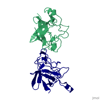
Interleukin-1 (IL-1) molecules are cytokines involved in the acute-phase | Interleukin-1 (IL-1) molecules are cytokines involved in the acute-phase response against infection and injury. Three naturally occurring IL-1 molecules are known, two agonists: IL-1 alpha and IL-1 beta, and one antagonist, the IL-1 receptor antagonist (IL-1ra). Although IL-1 action protects the organism by enhancing the response to pathogens, its overproduction can lead to pathology and has been implicated in disease states that include septic shock, rheumatoid arthritis, graft versus host disease and certain leukemias. The crystal structure of IL-1ra has been solved at 0.21-nm resolution by molecular replacement using the IL-1 beta structure as a search model. The crystals contain two independent IL-1ra molecules which are very similar. IL-1ra has the same fold as IL-1 alpha and IL-1 beta. The fold consists of twelve beta-strands which form a six-stranded beta-barrel, closed on one side by three beta-hairpin loops. Cys69 and Cys116 are linked via a disulfide bond and Pro53 has been built in the cis-conformation. Comparison of the IL-1ra structure with the IL-1 alpha and IL-1 beta structures present in the Protein Data Bank shows that a putative receptor interaction region, involving the N-terminus up to the beginning of strand beta 1 and the loops D and G, is very different in the three IL-1 molecules. Other putative interaction regions, as identified with mutagenesis studies, are structurally conserved and rigid, allowing precise and specific interactions with the IL-1 receptor. | ||
==Disease== | ==Disease== | ||
| Line 11: | Line 10: | ||
==About this Structure== | ==About this Structure== | ||
1ILR is a [http://en.wikipedia.org/wiki/Single_protein Single protein] structure of sequence from [http://en.wikipedia.org/wiki/Homo_sapiens Homo sapiens]. Full crystallographic information is available from [http:// | 1ILR is a [http://en.wikipedia.org/wiki/Single_protein Single protein] structure of sequence from [http://en.wikipedia.org/wiki/Homo_sapiens Homo sapiens]. Full crystallographic information is available from [http://oca.weizmann.ac.il/oca-bin/ocashort?id=1ILR OCA]. | ||
==Reference== | ==Reference== | ||
| Line 17: | Line 16: | ||
[[Category: Homo sapiens]] | [[Category: Homo sapiens]] | ||
[[Category: Single protein]] | [[Category: Single protein]] | ||
[[Category: Rondeau, J | [[Category: Rondeau, J M.]] | ||
[[Category: Schreuder, H | [[Category: Schreuder, H A.]] | ||
[[Category: Tardif, C.]] | [[Category: Tardif, C.]] | ||
[[Category: binding protein]] | [[Category: binding protein]] | ||
''Page seeded by [http:// | ''Page seeded by [http://oca.weizmann.ac.il/oca OCA ] on Thu Feb 21 13:13:10 2008'' | ||
Revision as of 14:13, 21 February 2008
|
CRYSTAL STRUCTURE OF THE INTERLEUKIN-1 RECEPTOR ANTAGONIST
OverviewOverview
Interleukin-1 (IL-1) molecules are cytokines involved in the acute-phase response against infection and injury. Three naturally occurring IL-1 molecules are known, two agonists: IL-1 alpha and IL-1 beta, and one antagonist, the IL-1 receptor antagonist (IL-1ra). Although IL-1 action protects the organism by enhancing the response to pathogens, its overproduction can lead to pathology and has been implicated in disease states that include septic shock, rheumatoid arthritis, graft versus host disease and certain leukemias. The crystal structure of IL-1ra has been solved at 0.21-nm resolution by molecular replacement using the IL-1 beta structure as a search model. The crystals contain two independent IL-1ra molecules which are very similar. IL-1ra has the same fold as IL-1 alpha and IL-1 beta. The fold consists of twelve beta-strands which form a six-stranded beta-barrel, closed on one side by three beta-hairpin loops. Cys69 and Cys116 are linked via a disulfide bond and Pro53 has been built in the cis-conformation. Comparison of the IL-1ra structure with the IL-1 alpha and IL-1 beta structures present in the Protein Data Bank shows that a putative receptor interaction region, involving the N-terminus up to the beginning of strand beta 1 and the loops D and G, is very different in the three IL-1 molecules. Other putative interaction regions, as identified with mutagenesis studies, are structurally conserved and rigid, allowing precise and specific interactions with the IL-1 receptor.
DiseaseDisease
Known diseases associated with this structure: Gastric cancer risk after H. pylori infection OMIM:[147679], Mental retardation, X-linked, 21/34 OMIM:[300206]
About this StructureAbout this Structure
1ILR is a Single protein structure of sequence from Homo sapiens. Full crystallographic information is available from OCA.
ReferenceReference
Refined crystal structure of the interleukin-1 receptor antagonist. Presence of a disulfide link and a cis-proline., Schreuder HA, Rondeau JM, Tardif C, Soffientini A, Sarubbi E, Akeson A, Bowlin TL, Yanofsky S, Barrett RW, Eur J Biochem. 1995 Feb 1;227(3):838-47. PMID:7867645
Page seeded by OCA on Thu Feb 21 13:13:10 2008