2ren: Difference between revisions
No edit summary |
No edit summary |
||
| Line 3: | Line 3: | ||
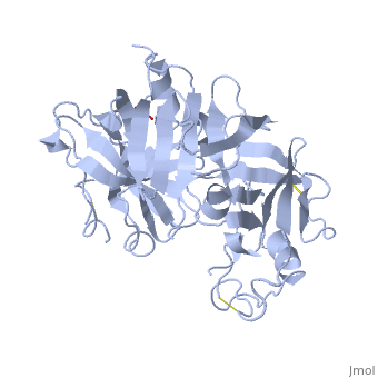
<StructureSection load='2ren' size='340' side='right'caption='[[2ren]], [[Resolution|resolution]] 2.50Å' scene=''> | <StructureSection load='2ren' size='340' side='right'caption='[[2ren]], [[Resolution|resolution]] 2.50Å' scene=''> | ||
== Structural highlights == | == Structural highlights == | ||
<table><tr><td colspan='2'>[[2ren]] is a 1 chain structure with sequence from [https://en.wikipedia.org/wiki/ | <table><tr><td colspan='2'>[[2ren]] is a 1 chain structure with sequence from [https://en.wikipedia.org/wiki/Homo_sapiens Homo sapiens]. Full crystallographic information is available from [http://oca.weizmann.ac.il/oca-bin/ocashort?id=2REN OCA]. For a <b>guided tour on the structure components</b> use [https://proteopedia.org/fgij/fg.htm?mol=2REN FirstGlance]. <br> | ||
</td></tr><tr id=' | </td></tr><tr id='method'><td class="sblockLbl"><b>[[Empirical_models|Method:]]</b></td><td class="sblockDat" id="methodDat">X-ray diffraction, [[Resolution|Resolution]] 2.5Å</td></tr> | ||
<tr id=' | <tr id='ligand'><td class="sblockLbl"><b>[[Ligand|Ligands:]]</b></td><td class="sblockDat" id="ligandDat"><scene name='pdbligand=NAG:N-ACETYL-D-GLUCOSAMINE'>NAG</scene></td></tr> | ||
<tr id='resources'><td class="sblockLbl"><b>Resources:</b></td><td class="sblockDat"><span class='plainlinks'>[https://proteopedia.org/fgij/fg.htm?mol=2ren FirstGlance], [http://oca.weizmann.ac.il/oca-bin/ocaids?id=2ren OCA], [https://pdbe.org/2ren PDBe], [https://www.rcsb.org/pdb/explore.do?structureId=2ren RCSB], [https://www.ebi.ac.uk/pdbsum/2ren PDBsum], [https://prosat.h-its.org/prosat/prosatexe?pdbcode=2ren ProSAT]</span></td></tr> | <tr id='resources'><td class="sblockLbl"><b>Resources:</b></td><td class="sblockDat"><span class='plainlinks'>[https://proteopedia.org/fgij/fg.htm?mol=2ren FirstGlance], [http://oca.weizmann.ac.il/oca-bin/ocaids?id=2ren OCA], [https://pdbe.org/2ren PDBe], [https://www.rcsb.org/pdb/explore.do?structureId=2ren RCSB], [https://www.ebi.ac.uk/pdbsum/2ren PDBsum], [https://prosat.h-its.org/prosat/prosatexe?pdbcode=2ren ProSAT]</span></td></tr> | ||
</table> | </table> | ||
== Disease == | == Disease == | ||
[https://www.uniprot.org/uniprot/RENI_HUMAN RENI_HUMAN] Defects in REN are a cause of renal tubular dysgenesis (RTD) [MIM:[https://omim.org/entry/267430 267430]. RTD is an autosomal recessive severe disorder of renal tubular development characterized by persistent fetal anuria and perinatal death, probably due to pulmonary hypoplasia from early-onset oligohydramnios (the Potter phenotype).<ref>PMID:16116425</ref> Defects in REN are the cause of familial juvenile hyperuricemic nephropathy type 2 (HNFJ2) [MIM:[https://omim.org/entry/613092 613092]. It is a renal disease characterized by juvenile onset of hyperuricemia, slowly progressive renal failure and anemia.<ref>PMID:19664745</ref> | |||
== Function == | == Function == | ||
[https://www.uniprot.org/uniprot/RENI_HUMAN RENI_HUMAN] Renin is a highly specific endopeptidase, whose only known function is to generate angiotensin I from angiotensinogen in the plasma, initiating a cascade of reactions that produce an elevation of blood pressure and increased sodium retention by the kidney. | |||
== Evolutionary Conservation == | == Evolutionary Conservation == | ||
[[Image:Consurf_key_small.gif|200px|right]] | [[Image:Consurf_key_small.gif|200px|right]] | ||
| Line 17: | Line 17: | ||
<jmolCheckbox> | <jmolCheckbox> | ||
<scriptWhenChecked>; select protein; define ~consurf_to_do selected; consurf_initial_scene = true; script "/wiki/ConSurf/re/2ren_consurf.spt"</scriptWhenChecked> | <scriptWhenChecked>; select protein; define ~consurf_to_do selected; consurf_initial_scene = true; script "/wiki/ConSurf/re/2ren_consurf.spt"</scriptWhenChecked> | ||
<scriptWhenUnchecked>script /wiki/extensions/Proteopedia/spt/ | <scriptWhenUnchecked>script /wiki/extensions/Proteopedia/spt/initialview03.spt</scriptWhenUnchecked> | ||
<text>to colour the structure by Evolutionary Conservation</text> | <text>to colour the structure by Evolutionary Conservation</text> | ||
</jmolCheckbox> | </jmolCheckbox> | ||
| Line 38: | Line 38: | ||
__TOC__ | __TOC__ | ||
</StructureSection> | </StructureSection> | ||
[[Category: | [[Category: Homo sapiens]] | ||
[[Category: Large Structures]] | [[Category: Large Structures]] | ||
[[Category: James MNG]] | |||
[[Category: James | [[Category: Sielecki AR]] | ||
[[Category: Sielecki | |||
Latest revision as of 12:29, 6 November 2024
STRUCTURE OF RECOMBINANT HUMAN RENIN, A TARGET FOR CARDIOVASCULAR-ACTIVE DRUGS, AT 2.5 ANGSTROMS RESOLUTIONSTRUCTURE OF RECOMBINANT HUMAN RENIN, A TARGET FOR CARDIOVASCULAR-ACTIVE DRUGS, AT 2.5 ANGSTROMS RESOLUTION
Structural highlights
DiseaseRENI_HUMAN Defects in REN are a cause of renal tubular dysgenesis (RTD) [MIM:267430. RTD is an autosomal recessive severe disorder of renal tubular development characterized by persistent fetal anuria and perinatal death, probably due to pulmonary hypoplasia from early-onset oligohydramnios (the Potter phenotype).[1] Defects in REN are the cause of familial juvenile hyperuricemic nephropathy type 2 (HNFJ2) [MIM:613092. It is a renal disease characterized by juvenile onset of hyperuricemia, slowly progressive renal failure and anemia.[2] FunctionRENI_HUMAN Renin is a highly specific endopeptidase, whose only known function is to generate angiotensin I from angiotensinogen in the plasma, initiating a cascade of reactions that produce an elevation of blood pressure and increased sodium retention by the kidney. Evolutionary Conservation Check, as determined by ConSurfDB. You may read the explanation of the method and the full data available from ConSurf. Publication Abstract from PubMedThe x-ray crystal structure of recombinant human renin has been determined. Molecular dynamics techniques that included crystallographic data as a restraint were used to improve an initial model based on porcine pepsinogen. The present agreement factor for data from 8.0 to 2.5 angstroms (A) is 0.236. Some of the surface loops are poorly determined, and these disordered regions border a 30 A wide solvent channel. Comparison of renin with other aspartyl proteinases shows that, although the structural cores and active sites are highly conserved, surface residues, some of which are critical for specificity, vary greatly (up to 10A). Knowledge of the actual structure, as opposed to the use of models based on related enzymes, should facilitate the design of renin inhibitors. Structure of recombinant human renin, a target for cardiovascular-active drugs, at 2.5 A resolution.,Sielecki AR, Hayakawa K, Fujinaga M, Murphy ME, Fraser M, Muir AK, Carilli CT, Lewicki JA, Baxter JD, James MN Science. 1989 Mar 10;243(4896):1346-51. PMID:2493678[3] From MEDLINE®/PubMed®, a database of the U.S. National Library of Medicine. See AlsoReferences
|
| ||||||||||||||||||