1i10: Difference between revisions
No edit summary |
No edit summary |
||
| Line 1: | Line 1: | ||
[[Image:1i10.gif|left|200px]] | [[Image:1i10.gif|left|200px]] | ||
<!-- | |||
The line below this paragraph, containing "STRUCTURE_1i10", creates the "Structure Box" on the page. | |||
You may change the PDB parameter (which sets the PDB file loaded into the applet) | |||
or the SCENE parameter (which sets the initial scene displayed when the page is loaded), | |||
or leave the SCENE parameter empty for the default display. | |||
| | --> | ||
| | {{STRUCTURE_1i10| PDB=1i10 | SCENE= }} | ||
}} | |||
'''HUMAN MUSCLE L-LACTATE DEHYDROGENASE M CHAIN, TERNARY COMPLEX WITH NADH AND OXAMATE''' | '''HUMAN MUSCLE L-LACTATE DEHYDROGENASE M CHAIN, TERNARY COMPLEX WITH NADH AND OXAMATE''' | ||
| Line 34: | Line 31: | ||
[[Category: Sessions, R B.]] | [[Category: Sessions, R B.]] | ||
[[Category: Winter, V J.]] | [[Category: Winter, V J.]] | ||
[[Category: | [[Category: Dehydrogenase]] | ||
[[Category: | [[Category: Rossman fold]] | ||
''Page seeded by [http://oca.weizmann.ac.il/oca OCA ] on Fri May 2 19:26:47 2008'' | |||
''Page seeded by [http://oca.weizmann.ac.il/oca OCA ] on | |||
Revision as of 19:26, 2 May 2008

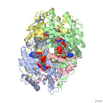
HUMAN MUSCLE L-LACTATE DEHYDROGENASE M CHAIN, TERNARY COMPLEX WITH NADH AND OXAMATE
OverviewOverview
Lactate dehydrogenase (LDH) interconverts pyruvate and lactate with concomitant interconversion of NADH and NAD(+). Although crystal structures of a variety of LDH have previously been described, a notable absence has been any of the three known human forms of this glycolytic enzyme. We have now determined the crystal structures of two isoforms of human LDH-the M form, predominantly found in muscle; and the H form, found mainly in cardiac muscle. Both structures have been crystallized as ternary complexes in the presence of the NADH cofactor and oxamate, a substrate-like inhibitor. Although each of these isoforms has different kinetic properties, the domain structure, subunit association, and active-site regions are indistinguishable between the two structures. The pK(a) that governs the K(M) for pyruvate for the two isozymes is found to differ by about 0.94 pH units, consistent with variation in pK(a) of the active-site histidine. The close similarity of these crystal structures suggests the distinctive activity of these enzyme isoforms is likely to result directly from variation of charged surface residues peripheral to the active site, a hypothesis supported by electrostatic calculations based on each structure. Proteins 2001;43:175-185.
DiseaseDisease
Known disease associated with this structure: Exertional myoglobinuria due to deficiency of LDH-A OMIM:[150000]
About this StructureAbout this Structure
1I10 is a Single protein structure of sequence from Homo sapiens. Full crystallographic information is available from OCA.
ReferenceReference
Structural basis for altered activity of M- and H-isozyme forms of human lactate dehydrogenase., Read JA, Winter VJ, Eszes CM, Sessions RB, Brady RL, Proteins. 2001 May 1;43(2):175-85. PMID:11276087 Page seeded by OCA on Fri May 2 19:26:47 2008