2cfp: Difference between revisions
No edit summary |
No edit summary |
||
| Line 1: | Line 1: | ||
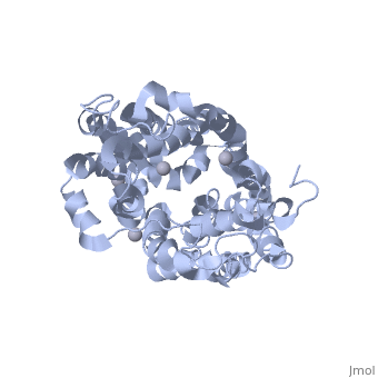
==SUGAR FREE LACTOSE PERMEASE AT ACIDIC PH== | |||
=== | <StructureSection load='2cfp' size='340' side='right' caption='[[2cfp]], [[Resolution|resolution]] 3.30Å' scene=''> | ||
== Structural highlights == | |||
<table><tr><td colspan='2'>[[2cfp]] is a 1 chain structure with sequence from [http://en.wikipedia.org/wiki/Escherichia_coli Escherichia coli]. Full crystallographic information is available from [http://oca.weizmann.ac.il/oca-bin/ocashort?id=2CFP OCA]. For a <b>guided tour on the structure components</b> use [http://oca.weizmann.ac.il/oca-docs/fgij/fg.htm?mol=2CFP FirstGlance]. <br> | |||
</td></tr><tr><td class="sblockLbl"><b>[[Ligand|Ligands:]]</b></td><td class="sblockDat"><scene name='pdbligand=HG:MERCURY+(II)+ION'>HG</scene><br> | |||
<tr><td class="sblockLbl"><b>[[Related_structure|Related:]]</b></td><td class="sblockDat">[[1m2u|1m2u]], [[1pv6|1pv6]], [[1pv7|1pv7]], [[2cfq|2cfq]]</td></tr> | |||
<tr><td class="sblockLbl"><b>Resources:</b></td><td class="sblockDat"><span class='plainlinks'>[http://oca.weizmann.ac.il/oca-docs/fgij/fg.htm?mol=2cfp FirstGlance], [http://oca.weizmann.ac.il/oca-bin/ocaids?id=2cfp OCA], [http://www.rcsb.org/pdb/explore.do?structureId=2cfp RCSB], [http://www.ebi.ac.uk/pdbsum/2cfp PDBsum]</span></td></tr> | |||
<table> | |||
== Evolutionary Conservation == | |||
[[Image:Consurf_key_small.gif|200px|right]] | |||
Check<jmol> | |||
<jmolCheckbox> | |||
<scriptWhenChecked>select protein; define ~consurf_to_do selected; consurf_initial_scene = true; script "/wiki/ConSurf/cf/2cfp_consurf.spt"</scriptWhenChecked> | |||
<scriptWhenUnchecked>script /wiki/extensions/Proteopedia/spt/initialview01.spt</scriptWhenUnchecked> | |||
<text>to colour the structure by Evolutionary Conservation</text> | |||
</jmolCheckbox> | |||
</jmol>, as determined by [http://consurfdb.tau.ac.il/ ConSurfDB]. You may read the [[Conservation%2C_Evolutionary|explanation]] of the method and the full data available from [http://bental.tau.ac.il/new_ConSurfDB/chain_selection.php?pdb_ID=2ata ConSurf]. | |||
<div style="clear:both"></div> | |||
<div style="background-color:#fffaf0;"> | |||
== Publication Abstract from PubMed == | |||
Cation-coupled active transport is an essential cellular process found ubiquitously in all living organisms. Here, we present two novel ligand-free X-ray structures of the lactose permease (LacY) of Escherichia coli determined at acidic and neutral pH, and propose a model for the mechanism of coupling between lactose and H+ translocation. No sugar-binding site is observed in the absence of ligand, and deprotonation of the key residue Glu269 is associated with ligand binding. Thus, substrate induces formation of the sugar-binding site, as well as the initial step in H+ transduction. | |||
Structural evidence for induced fit and a mechanism for sugar/H+ symport in LacY.,Mirza O, Guan L, Verner G, Iwata S, Kaback HR EMBO J. 2006 Mar 22;25(6):1177-83. Epub 2006 Mar 9. PMID:16525509<ref>PMID:16525509</ref> | |||
From MEDLINE®/PubMed®, a database of the U.S. National Library of Medicine.<br> | |||
</div> | |||
==See Also== | ==See Also== | ||
*[[Lactose Permease|Lactose Permease]] | *[[Lactose Permease|Lactose Permease]] | ||
*[[Major Facilitators|Major Facilitators]] | *[[Major Facilitators|Major Facilitators]] | ||
== References == | |||
== | <references/> | ||
< | __TOC__ | ||
</StructureSection> | |||
[[Category: Escherichia coli]] | [[Category: Escherichia coli]] | ||
[[Category: Guan, L.]] | [[Category: Guan, L.]] | ||
Revision as of 05:17, 30 September 2014
SUGAR FREE LACTOSE PERMEASE AT ACIDIC PHSUGAR FREE LACTOSE PERMEASE AT ACIDIC PH
Structural highlights
Evolutionary Conservation Check, as determined by ConSurfDB. You may read the explanation of the method and the full data available from ConSurf. Publication Abstract from PubMedCation-coupled active transport is an essential cellular process found ubiquitously in all living organisms. Here, we present two novel ligand-free X-ray structures of the lactose permease (LacY) of Escherichia coli determined at acidic and neutral pH, and propose a model for the mechanism of coupling between lactose and H+ translocation. No sugar-binding site is observed in the absence of ligand, and deprotonation of the key residue Glu269 is associated with ligand binding. Thus, substrate induces formation of the sugar-binding site, as well as the initial step in H+ transduction. Structural evidence for induced fit and a mechanism for sugar/H+ symport in LacY.,Mirza O, Guan L, Verner G, Iwata S, Kaback HR EMBO J. 2006 Mar 22;25(6):1177-83. Epub 2006 Mar 9. PMID:16525509[1] From MEDLINE®/PubMed®, a database of the U.S. National Library of Medicine. See AlsoReferences |
| ||||||||||||||||||