2bat: Difference between revisions
No edit summary |
No edit summary |
||
| Line 1: | Line 1: | ||
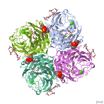
==THE STRUCTURE OF THE COMPLEX BETWEEN INFLUENZA VIRUS NEURAMINIDASE AND SIALIC ACID, THE VIRAL RECEPTOR== | |||
<StructureSection load='2bat' size='340' side='right' caption='[[2bat]], [[Resolution|resolution]] 2.00Å' scene=''> | |||
== Structural highlights == | |||
<table><tr><td colspan='2'>[[2bat]] is a 1 chain structure with sequence from [http://en.wikipedia.org/wiki/Viruses Viruses]. The May 2009 RCSB PDB [http://pdb.rcsb.org/pdb/static.do?p=education_discussion/molecule_of_the_month/index.html Molecule of the Month] feature on ''Influenza Neuraminidase'' by David Goodsell is [http://dx.doi.org/10.2210/rcsb_pdb/mom_2009_5 10.2210/rcsb_pdb/mom_2009_5]. Full crystallographic information is available from [http://oca.weizmann.ac.il/oca-bin/ocashort?id=2BAT OCA]. For a <b>guided tour on the structure components</b> use [http://oca.weizmann.ac.il/oca-docs/fgij/fg.htm?mol=2BAT FirstGlance]. <br> | |||
</td></tr><tr><td class="sblockLbl"><b>[[Ligand|Ligands:]]</b></td><td class="sblockDat"><scene name='pdbligand=BMA:BETA-D-MANNOSE'>BMA</scene>, <scene name='pdbligand=CA:CALCIUM+ION'>CA</scene>, <scene name='pdbligand=NAG:N-ACETYL-D-GLUCOSAMINE'>NAG</scene>, <scene name='pdbligand=SIA:O-SIALIC+ACID'>SIA</scene>, <scene name='pdbligand=FUL:BETA-L-FUCOSE'>FUL</scene>, <scene name='pdbligand=MAN:ALPHA-D-MANNOSE'>MAN</scene><br> | |||
<tr><td class="sblockLbl"><b>[[Non-Standard_Residue|NonStd Res:]]</b></td><td class="sblockDat"><scene name='pdbligand=NGK:2-(ACETYLAMINO)-2-DEOXY-4-O-SULFO-ALPHA-D-GALACTOPYRANOSE'>NGK</scene></td></tr> | |||
<tr><td class="sblockLbl"><b>[[Related_structure|Related:]]</b></td><td class="sblockDat">[[1nn2|1nn2]]</td></tr> | |||
<tr><td class="sblockLbl"><b>Activity:</b></td><td class="sblockDat"><span class='plainlinks'>[http://en.wikipedia.org/wiki/Exo-alpha-sialidase Exo-alpha-sialidase], with EC number [http://www.brenda-enzymes.info/php/result_flat.php4?ecno=3.2.1.18 3.2.1.18] </span></td></tr> | |||
<tr><td class="sblockLbl"><b>Resources:</b></td><td class="sblockDat"><span class='plainlinks'>[http://oca.weizmann.ac.il/oca-docs/fgij/fg.htm?mol=2bat FirstGlance], [http://oca.weizmann.ac.il/oca-bin/ocaids?id=2bat OCA], [http://www.rcsb.org/pdb/explore.do?structureId=2bat RCSB], [http://www.ebi.ac.uk/pdbsum/2bat PDBsum]</span></td></tr> | |||
<table> | |||
== Evolutionary Conservation == | |||
[[Image:Consurf_key_small.gif|200px|right]] | |||
Check<jmol> | |||
<jmolCheckbox> | |||
<scriptWhenChecked>select protein; define ~consurf_to_do selected; consurf_initial_scene = true; script "/wiki/ConSurf/ba/2bat_consurf.spt"</scriptWhenChecked> | |||
<scriptWhenUnchecked>script /wiki/extensions/Proteopedia/spt/initialview01.spt</scriptWhenUnchecked> | |||
<text>to colour the structure by Evolutionary Conservation</text> | |||
</jmolCheckbox> | |||
</jmol>, as determined by [http://consurfdb.tau.ac.il/ ConSurfDB]. You may read the [[Conservation%2C_Evolutionary|explanation]] of the method and the full data available from [http://bental.tau.ac.il/new_ConSurfDB/chain_selection.php?pdb_ID=2ata ConSurf]. | |||
<div style="clear:both"></div> | |||
<div style="background-color:#fffaf0;"> | |||
== Publication Abstract from PubMed == | |||
Crystallographic studies of neuraminidase-sialic acid complexes indicate that sialic acid is distorted on binding the enzyme. Three arginine residues on the enzyme interact with the carboxylate group of the sugar which is observed to be equatorial to the saccharide ring as a consequence of its distorted geometry. The glycosidic oxygen is positioned within hydrogen-bonding distance of Asp-151, implicating this residue in catalysis. | |||
The structure of the complex between influenza virus neuraminidase and sialic acid, the viral receptor.,Varghese JN, McKimm-Breschkin JL, Caldwell JB, Kortt AA, Colman PM Proteins. 1992 Nov;14(3):327-32. PMID:1438172<ref>PMID:1438172</ref> | |||
From MEDLINE®/PubMed®, a database of the U.S. National Library of Medicine.<br> | |||
</div> | |||
==See Also== | ==See Also== | ||
*[[Neuraminidase|Neuraminidase]] | *[[Neuraminidase|Neuraminidase]] | ||
*[[Teaching Scenes%2C Tutorials%2C and Educators' Pages|Teaching Scenes%2C Tutorials%2C and Educators' Pages]] | *[[Teaching Scenes%2C Tutorials%2C and Educators' Pages|Teaching Scenes%2C Tutorials%2C and Educators' Pages]] | ||
== References == | |||
== | <references/> | ||
< | __TOC__ | ||
</StructureSection> | |||
[[Category: Exo-alpha-sialidase]] | [[Category: Exo-alpha-sialidase]] | ||
[[Category: Influenza Neuraminidase]] | [[Category: Influenza Neuraminidase]] | ||
Revision as of 04:15, 30 September 2014
THE STRUCTURE OF THE COMPLEX BETWEEN INFLUENZA VIRUS NEURAMINIDASE AND SIALIC ACID, THE VIRAL RECEPTORTHE STRUCTURE OF THE COMPLEX BETWEEN INFLUENZA VIRUS NEURAMINIDASE AND SIALIC ACID, THE VIRAL RECEPTOR
Structural highlights
Evolutionary Conservation Check, as determined by ConSurfDB. You may read the explanation of the method and the full data available from ConSurf. Publication Abstract from PubMedCrystallographic studies of neuraminidase-sialic acid complexes indicate that sialic acid is distorted on binding the enzyme. Three arginine residues on the enzyme interact with the carboxylate group of the sugar which is observed to be equatorial to the saccharide ring as a consequence of its distorted geometry. The glycosidic oxygen is positioned within hydrogen-bonding distance of Asp-151, implicating this residue in catalysis. The structure of the complex between influenza virus neuraminidase and sialic acid, the viral receptor.,Varghese JN, McKimm-Breschkin JL, Caldwell JB, Kortt AA, Colman PM Proteins. 1992 Nov;14(3):327-32. PMID:1438172[1] From MEDLINE®/PubMed®, a database of the U.S. National Library of Medicine. See AlsoReferences
|
| ||||||||||||||||||||||