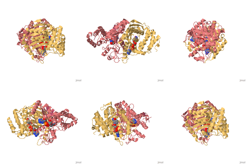
2x06: Difference between revisions
m Protected "2x06" [edit=sysop:move=sysop] |
No edit summary |
||
| Line 1: | Line 1: | ||
[[Image: | ==SULFOLACTATE DEHYDROGENASE FROM METHANOCALDOCOCCUS JANNASCHII== | ||
<StructureSection load='2x06' size='340' side='right' caption='[[2x06]], [[Resolution|resolution]] 2.50Å' scene=''> | |||
== Structural highlights == | |||
<table><tr><td colspan='2'>[[2x06]] is a 8 chain structure with sequence from [http://en.wikipedia.org/wiki/Methanocaldococcus_jannaschii Methanocaldococcus jannaschii]. This structure supersedes the now removed PDB entry [http://oca.weizmann.ac.il/oca-bin/send-pdb?obs=1&id=1rfm 1rfm]. Full crystallographic information is available from [http://oca.weizmann.ac.il/oca-bin/ocashort?id=2X06 OCA]. <br> | |||
</td></tr><tr><td class="sblockLbl"><b>[[Ligand|Ligands:]]</b></td><td class="sblockDat"><scene name='pdbligand=NAD:NICOTINAMIDE-ADENINE-DINUCLEOTIDE'>NAD</scene><br> | |||
<tr><td class="sblockLbl"><b>[[Non-Standard_Residue|NonStd Res:]]</b></td><td class="sblockDat"><scene name='pdbligand=MSE:SELENOMETHIONINE'>MSE</scene></td></tr> | |||
<tr><td class="sblockLbl"><b>Activity:</b></td><td class="sblockDat"><span class='plainlinks'>[http://en.wikipedia.org/wiki/Glucokinase Glucokinase], with EC number [http://www.brenda-enzymes.info/php/result_flat.php4?ecno=2.7.1.2 2.7.1.2] </span></td></tr> | |||
<tr><td class="sblockLbl"><b>Resources:</b></td><td class="sblockDat"><span class='plainlinks'>[http://oca.weizmann.ac.il/oca-docs/fgij/fg.htm?mol=2x06 FirstGlance], [http://oca.weizmann.ac.il/oca-bin/ocaids?id=2x06 OCA], [http://www.rcsb.org/pdb/explore.do?structureId=2x06 RCSB], [http://www.ebi.ac.uk/pdbsum/2x06 PDBsum]</span></td></tr> | |||
<table> | |||
== Evolutionary Conservation == | |||
[[Image:Consurf_key_small.gif|200px|right]] | |||
Check<jmol> | |||
<jmolCheckbox> | |||
<scriptWhenChecked>select protein; define ~consurf_to_do selected; consurf_initial_scene = true; script "/wiki/ConSurf/x0/2x06_consurf.spt"</scriptWhenChecked> | |||
<scriptWhenUnchecked>script /wiki/extensions/Proteopedia/spt/initialview01.spt</scriptWhenUnchecked> | |||
<text>to colour the structure by Evolutionary Conservation</text> | |||
</jmolCheckbox> | |||
</jmol>, as determined by [http://consurfdb.tau.ac.il/ ConSurfDB]. You may read the [[Conservation%2C_Evolutionary|explanation]] of the method and the full data available from [http://bental.tau.ac.il/new_ConSurfDB/chain_selection.php?pdb_ID=2ata ConSurf]. | |||
<div style="clear:both"></div> | |||
<div style="background-color:#fffaf0;"> | |||
== Publication Abstract from PubMed == | |||
The crystal structure of the sulfolactate dehydrogenase from the hyperthermophilic and methanogenic archaeon Methanocaldococcus jannaschii was solved at 2.5 A resolution (PDB id. 1RFM). The asymmetric unit contains a tetramer of tight dimers. This structure, complexed with NADH, does not contain a cofactor-binding domain with 'Rossmann-fold' topology. Instead, the tertiary and quaternary structures indicate a novel fold. The NADH is bound in an extended conformation in each active site, in a manner that explains the pro-S specificity. Cofactor binding involves residues belonging to both subunits within the tight dimers, which are therefore the smallest enzymatically active units. The protein was found to be a homodimer in solution by size-exclusion chromatography, analytical ultracentrifugation and small-angle neutron scattering. Various compounds were tested as putative substrates. The results indicate the existence of a substrate discrimination mechanism, which involves electrostatic interactions. Based on sequence homology and phylogenetic analyses, several other enzymes were classified as belonging to this novel family of homologous (S)-2-hydroxyacid dehydrogenases. | |||
Methanoarchaeal sulfolactate dehydrogenase: prototype of a new family of NADH-dependent enzymes.,Irimia A, Madern D, Zaccai G, Vellieux FM EMBO J. 2004 Mar 24;23(6):1234-44. Epub 2004 Mar 11. PMID:15014443<ref>PMID:15014443</ref> | |||
From MEDLINE®/PubMed®, a database of the U.S. National Library of Medicine.<br> | |||
</div> | |||
== References == | |||
<references/> | |||
__TOC__ | |||
</StructureSection> | |||
== | |||
< | |||
[[Category: Methanocaldococcus jannaschii]] | [[Category: Methanocaldococcus jannaschii]] | ||
[[Category: Irimia, A.]] | [[Category: Irimia, A.]] | ||
Revision as of 10:43, 14 May 2014
SULFOLACTATE DEHYDROGENASE FROM METHANOCALDOCOCCUS JANNASCHIISULFOLACTATE DEHYDROGENASE FROM METHANOCALDOCOCCUS JANNASCHII
Structural highlights
Evolutionary Conservation Check, as determined by ConSurfDB. You may read the explanation of the method and the full data available from ConSurf. Publication Abstract from PubMedThe crystal structure of the sulfolactate dehydrogenase from the hyperthermophilic and methanogenic archaeon Methanocaldococcus jannaschii was solved at 2.5 A resolution (PDB id. 1RFM). The asymmetric unit contains a tetramer of tight dimers. This structure, complexed with NADH, does not contain a cofactor-binding domain with 'Rossmann-fold' topology. Instead, the tertiary and quaternary structures indicate a novel fold. The NADH is bound in an extended conformation in each active site, in a manner that explains the pro-S specificity. Cofactor binding involves residues belonging to both subunits within the tight dimers, which are therefore the smallest enzymatically active units. The protein was found to be a homodimer in solution by size-exclusion chromatography, analytical ultracentrifugation and small-angle neutron scattering. Various compounds were tested as putative substrates. The results indicate the existence of a substrate discrimination mechanism, which involves electrostatic interactions. Based on sequence homology and phylogenetic analyses, several other enzymes were classified as belonging to this novel family of homologous (S)-2-hydroxyacid dehydrogenases. Methanoarchaeal sulfolactate dehydrogenase: prototype of a new family of NADH-dependent enzymes.,Irimia A, Madern D, Zaccai G, Vellieux FM EMBO J. 2004 Mar 24;23(6):1234-44. Epub 2004 Mar 11. PMID:15014443[1] From MEDLINE®/PubMed®, a database of the U.S. National Library of Medicine. References
|
| ||||||||||||||||||||
