1ilr: Difference between revisions
No edit summary |
No edit summary |
||
| (13 intermediate revisions by the same user not shown) | |||
| Line 1: | Line 1: | ||
< | ==CRYSTAL STRUCTURE OF THE INTERLEUKIN-1 RECEPTOR ANTAGONIST== | ||
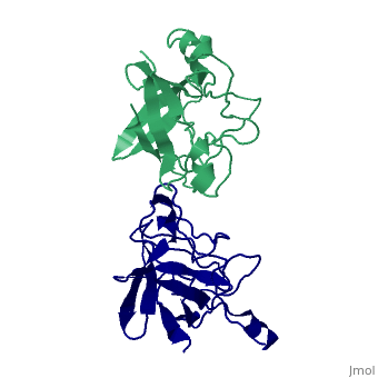
<StructureSection load='1ilr' size='340' side='right'caption='[[1ilr]], [[Resolution|resolution]] 2.10Å' scene=''> | |||
== Structural highlights == | |||
or | <table><tr><td colspan='2'>[[1ilr]] is a 2 chain structure with sequence from [https://en.wikipedia.org/wiki/Homo_sapiens Homo sapiens]. Full crystallographic information is available from [http://oca.weizmann.ac.il/oca-bin/ocashort?id=1ILR OCA]. For a <b>guided tour on the structure components</b> use [https://proteopedia.org/fgij/fg.htm?mol=1ILR FirstGlance]. <br> | ||
</td></tr><tr id='method'><td class="sblockLbl"><b>[[Empirical_models|Method:]]</b></td><td class="sblockDat" id="methodDat">X-ray diffraction, [[Resolution|Resolution]] 2.1Å</td></tr> | |||
<tr id='resources'><td class="sblockLbl"><b>Resources:</b></td><td class="sblockDat"><span class='plainlinks'>[https://proteopedia.org/fgij/fg.htm?mol=1ilr FirstGlance], [http://oca.weizmann.ac.il/oca-bin/ocaids?id=1ilr OCA], [https://pdbe.org/1ilr PDBe], [https://www.rcsb.org/pdb/explore.do?structureId=1ilr RCSB], [https://www.ebi.ac.uk/pdbsum/1ilr PDBsum], [https://prosat.h-its.org/prosat/prosatexe?pdbcode=1ilr ProSAT]</span></td></tr> | |||
</table> | |||
== Disease == | |||
[https://www.uniprot.org/uniprot/IL1RA_HUMAN IL1RA_HUMAN] Genetic variation in IL1RN is associated with susceptibility to microvascular complications of diabetes type 4 (MVCD4) [MIM:[https://omim.org/entry/612628 612628]. These are pathological conditions that develop in numerous tissues and organs as a consequence of diabetes mellitus. They include diabetic retinopathy, diabetic nephropathy leading to end-stage renal disease, and diabetic neuropathy. Diabetic retinopathy remains the major cause of new-onset blindness among diabetic adults. It is characterized by vascular permeability and increased tissue ischemia and angiogenesis. Defects in IL1RN are the cause of interleukin 1 receptor antagonist deficiency (DIRA) [MIM:[https://omim.org/entry/612852 612852]; also known as deficiency of interleukin 1 receptor antagonist. Autoinflammatory diseases manifest inflammation without evidence of infection, high-titer autoantibodies, or autoreactive T-cells. DIRA is a rare, autosomal recessive, genetic autoinflammatory disease that results in sterile multifocal osteomyelitis (bone inflammation in multiple places), periostitis (inflammation of the membrane surrounding the bones), and pustulosis (due to skin inflammation) from birth.<ref>PMID:19494218</ref> | |||
== Function == | |||
[https://www.uniprot.org/uniprot/IL1RA_HUMAN IL1RA_HUMAN] Inhibits the activity of interleukin-1 by binding to receptor IL1R1 and preventing its association with the coreceptor IL1RAP for signaling. Has no interleukin-1 like activity. Binds functional interleukin-1 receptor IL1R1 with greater affinity than decoy receptor IL1R2; however, the physiological relevance of the latter association is unsure.<ref>PMID:7775431</ref> | |||
== Evolutionary Conservation == | |||
[[Image:Consurf_key_small.gif|200px|right]] | |||
Check<jmol> | |||
<jmolCheckbox> | |||
<scriptWhenChecked>; select protein; define ~consurf_to_do selected; consurf_initial_scene = true; script "/wiki/ConSurf/il/1ilr_consurf.spt"</scriptWhenChecked> | |||
<scriptWhenUnchecked>script /wiki/extensions/Proteopedia/spt/initialview03.spt</scriptWhenUnchecked> | |||
<text>to colour the structure by Evolutionary Conservation</text> | |||
</jmolCheckbox> | |||
</jmol>, as determined by [http://consurfdb.tau.ac.il/ ConSurfDB]. You may read the [[Conservation%2C_Evolutionary|explanation]] of the method and the full data available from [http://bental.tau.ac.il/new_ConSurfDB/main_output.php?pdb_ID=1ilr ConSurf]. | |||
<div style="clear:both"></div> | |||
<div style="background-color:#fffaf0;"> | |||
== Publication Abstract from PubMed == | |||
Interleukin-1 (IL-1) molecules are cytokines involved in the acute-phase response against infection and injury. Three naturally occurring IL-1 molecules are known, two agonists: IL-1 alpha and IL-1 beta, and one antagonist, the IL-1 receptor antagonist (IL-1ra). Although IL-1 action protects the organism by enhancing the response to pathogens, its overproduction can lead to pathology and has been implicated in disease states that include septic shock, rheumatoid arthritis, graft versus host disease and certain leukemias. The crystal structure of IL-1ra has been solved at 0.21-nm resolution by molecular replacement using the IL-1 beta structure as a search model. The crystals contain two independent IL-1ra molecules which are very similar. IL-1ra has the same fold as IL-1 alpha and IL-1 beta. The fold consists of twelve beta-strands which form a six-stranded beta-barrel, closed on one side by three beta-hairpin loops. Cys69 and Cys116 are linked via a disulfide bond and Pro53 has been built in the cis-conformation. Comparison of the IL-1ra structure with the IL-1 alpha and IL-1 beta structures present in the Protein Data Bank shows that a putative receptor interaction region, involving the N-terminus up to the beginning of strand beta 1 and the loops D and G, is very different in the three IL-1 molecules. Other putative interaction regions, as identified with mutagenesis studies, are structurally conserved and rigid, allowing precise and specific interactions with the IL-1 receptor. | |||
Refined crystal structure of the interleukin-1 receptor antagonist. Presence of a disulfide link and a cis-proline.,Schreuder HA, Rondeau JM, Tardif C, Soffientini A, Sarubbi E, Akeson A, Bowlin TL, Yanofsky S, Barrett RW Eur J Biochem. 1995 Feb 1;227(3):838-47. PMID:7867645<ref>PMID:7867645</ref> | |||
From MEDLINE®/PubMed®, a database of the U.S. National Library of Medicine.<br> | |||
</div> | |||
<div class="pdbe-citations 1ilr" style="background-color:#fffaf0;"></div> | |||
==See Also== | |||
*[[Interleukin receptor antagonist|Interleukin receptor antagonist]] | |||
== References == | |||
<references/> | |||
__TOC__ | |||
</StructureSection> | |||
== | |||
== | |||
[[Category: Homo sapiens]] | [[Category: Homo sapiens]] | ||
[[Category: | [[Category: Large Structures]] | ||
[[Category: Rondeau | [[Category: Rondeau J-M]] | ||
[[Category: Schreuder | [[Category: Schreuder HA]] | ||
[[Category: Tardif | [[Category: Tardif C]] | ||
Latest revision as of 09:46, 30 October 2024
CRYSTAL STRUCTURE OF THE INTERLEUKIN-1 RECEPTOR ANTAGONISTCRYSTAL STRUCTURE OF THE INTERLEUKIN-1 RECEPTOR ANTAGONIST
Structural highlights
DiseaseIL1RA_HUMAN Genetic variation in IL1RN is associated with susceptibility to microvascular complications of diabetes type 4 (MVCD4) [MIM:612628. These are pathological conditions that develop in numerous tissues and organs as a consequence of diabetes mellitus. They include diabetic retinopathy, diabetic nephropathy leading to end-stage renal disease, and diabetic neuropathy. Diabetic retinopathy remains the major cause of new-onset blindness among diabetic adults. It is characterized by vascular permeability and increased tissue ischemia and angiogenesis. Defects in IL1RN are the cause of interleukin 1 receptor antagonist deficiency (DIRA) [MIM:612852; also known as deficiency of interleukin 1 receptor antagonist. Autoinflammatory diseases manifest inflammation without evidence of infection, high-titer autoantibodies, or autoreactive T-cells. DIRA is a rare, autosomal recessive, genetic autoinflammatory disease that results in sterile multifocal osteomyelitis (bone inflammation in multiple places), periostitis (inflammation of the membrane surrounding the bones), and pustulosis (due to skin inflammation) from birth.[1] FunctionIL1RA_HUMAN Inhibits the activity of interleukin-1 by binding to receptor IL1R1 and preventing its association with the coreceptor IL1RAP for signaling. Has no interleukin-1 like activity. Binds functional interleukin-1 receptor IL1R1 with greater affinity than decoy receptor IL1R2; however, the physiological relevance of the latter association is unsure.[2] Evolutionary Conservation Check, as determined by ConSurfDB. You may read the explanation of the method and the full data available from ConSurf. Publication Abstract from PubMedInterleukin-1 (IL-1) molecules are cytokines involved in the acute-phase response against infection and injury. Three naturally occurring IL-1 molecules are known, two agonists: IL-1 alpha and IL-1 beta, and one antagonist, the IL-1 receptor antagonist (IL-1ra). Although IL-1 action protects the organism by enhancing the response to pathogens, its overproduction can lead to pathology and has been implicated in disease states that include septic shock, rheumatoid arthritis, graft versus host disease and certain leukemias. The crystal structure of IL-1ra has been solved at 0.21-nm resolution by molecular replacement using the IL-1 beta structure as a search model. The crystals contain two independent IL-1ra molecules which are very similar. IL-1ra has the same fold as IL-1 alpha and IL-1 beta. The fold consists of twelve beta-strands which form a six-stranded beta-barrel, closed on one side by three beta-hairpin loops. Cys69 and Cys116 are linked via a disulfide bond and Pro53 has been built in the cis-conformation. Comparison of the IL-1ra structure with the IL-1 alpha and IL-1 beta structures present in the Protein Data Bank shows that a putative receptor interaction region, involving the N-terminus up to the beginning of strand beta 1 and the loops D and G, is very different in the three IL-1 molecules. Other putative interaction regions, as identified with mutagenesis studies, are structurally conserved and rigid, allowing precise and specific interactions with the IL-1 receptor. Refined crystal structure of the interleukin-1 receptor antagonist. Presence of a disulfide link and a cis-proline.,Schreuder HA, Rondeau JM, Tardif C, Soffientini A, Sarubbi E, Akeson A, Bowlin TL, Yanofsky S, Barrett RW Eur J Biochem. 1995 Feb 1;227(3):838-47. PMID:7867645[3] From MEDLINE®/PubMed®, a database of the U.S. National Library of Medicine. See AlsoReferences
|
| ||||||||||||||||