IgA: Difference between revisions
No edit summary |
Michal Harel (talk | contribs) No edit summary |
||
| (4 intermediate revisions by 2 users not shown) | |||
| Line 1: | Line 1: | ||
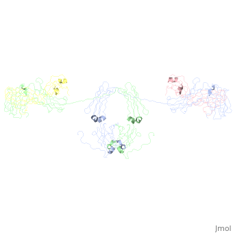
<StructureSection load='1iga' size='450' side='right' scene='' caption=''> | <StructureSection load='1iga' size='450' side='right' scene='' caption='Model of human Iga1 [[1iga]]'> | ||
== Introduction to IgA == | == Introduction to IgA == | ||
The most extensive surface in contact with the external environment is not our skin, but the epithelial lining of our gastrointestinal, respiratory, and urogenital tracts <ref name="seven">PMID:17428798</ref>. As a first line of defense in maintenance the integrity our mucosa, the immune system manufactures and secretes dimeric IgA to neutralize pathogenic organisms <ref name="five">PMID:15111057</ref> and exclude the entry of commensals at the mucosal border <ref name="nineseven">PMID:19079336</ref>. In the serum, IgA functions as a second line of defense against pathogens that may breech the epithelial boundary <ref name="five" />. The body produces more IgA than any other antibody isotype <ref name="nineseven"/>. In fact, IgA is the most abundant antibody in the body, further illustrating IgA's critical role in immunity <ref name="ten">PMID:10064707</ref>. | The most extensive surface in contact with the external environment is not our skin, but the epithelial lining of our gastrointestinal, respiratory, and urogenital tracts <ref name="seven">PMID:17428798</ref>. As a first line of defense in maintenance the integrity our mucosa, the immune system manufactures and secretes dimeric IgA to neutralize pathogenic organisms <ref name="five">PMID:15111057</ref> and exclude the entry of commensals at the mucosal border <ref name="nineseven">PMID:19079336</ref>. In the serum, IgA functions as a second line of defense against pathogens that may breech the epithelial boundary <ref name="five" />. The body produces more IgA than any other antibody isotype <ref name="nineseven"/>. In fact, IgA is the most abundant antibody in the body, further illustrating IgA's critical role in immunity <ref name="ten">PMID:10064707</ref>. | ||
| Line 16: | Line 17: | ||
'''Fab and Fc fragments''' | '''Fab and Fc fragments''' | ||
:Another common way of describing antibody structure is in terms of its Fab and Fc fragments. Each light chains are composed of 2 immunoglobulin domains: one variable domain | :Another common way of describing antibody structure is in terms of its Fab and Fc fragments. Each light chains are composed of 2 immunoglobulin domains: one variable domain and one constant domain. Heavy chains composed of 4 Ig domains: one V-type and 3 C-type, named CH1 - CH3. A linking hinge region separates the CH2 and CH3 domains. Proteolytic cleavage at the hinge region by the protease papain, or a similar protease, yields 2 Fab fragments and 1 Fc fragment. Each <scene name='Rebecca_Martin/Sandbox1/Fab_ex/1'>Fab fragment</scene> contains 2 variable domains, one from the heavy chain and one from the light chain, and 2 constant domains one from the light chain and the Ch1 domain from the heavy chain. The <scene name='Rebecca_Martin/Sandbox1/Fc/1'>Fc fragment</scene> Fc fragment contains 4 constant domains: the Ch2 and Ch3 domains from each of the heavy chains. Since the variable portions determine antigen specificity, the Fab fragments are generally thought of as the antigen-binding portion. The Fc fragment is important in binding various receptors, many of which are isotype specific and are named after the isotype of the ligand, i.e. FcαR binds the Fc portion of IgA. | ||
'''Immunoglobulin domains''' | '''Immunoglobulin domains''' | ||
| Line 155: | Line 156: | ||
:Crystallographic structure will yield further insights into the structure of IgA, the interactions between IgA and other molecules. | :Crystallographic structure will yield further insights into the structure of IgA, the interactions between IgA and other molecules. | ||
</StructureSection> | |||
__NOTOC__ | |||
== Links == | == Links == | ||
=== IgA === | === IgA === | ||
Latest revision as of 12:23, 11 July 2013
Introduction to IgAThe most extensive surface in contact with the external environment is not our skin, but the epithelial lining of our gastrointestinal, respiratory, and urogenital tracts [1]. As a first line of defense in maintenance the integrity our mucosa, the immune system manufactures and secretes dimeric IgA to neutralize pathogenic organisms [2] and exclude the entry of commensals at the mucosal border [3]. In the serum, IgA functions as a second line of defense against pathogens that may breech the epithelial boundary [2]. The body produces more IgA than any other antibody isotype [3]. In fact, IgA is the most abundant antibody in the body, further illustrating IgA's critical role in immunity [4].
At least two isotypes exist, termed IgA1 and IgA2. IgA2 can further be categorized into 2 allotypes: IgA2 m(1) and IgA2 m(2). While IgA2 is found in most mammalian species, IgA1 is found only in higher apes. An approximately equal ratio of secretory IgA1 (sIgA1) to secretory IgA2 (sIgA2) reside at the mucosal surface, with the exception of the colon, where the majority is sIgA2 [5]. In the serum, about 90% of the IgA is monomeric IgA1 [4]. While both isoforms are able to bind polysaccharide, IgA1 preferentially binds protein antigen, while IgA2 preferentially binds lipopolysaccharide lipid A.
The receptors for IgA include the Fcα Receptor (FcαRI; CD89) and the polyimmunologlobulin receptor (pIgR). When binding to FcαRI results in the dimerization, the consequent signaling results in effector functions, including respiratory burst, mucosal surface, phagocytosis, and eosinophil degranulation. Binding to the pIgR results in transocytosis and IgA secretion [2]. Unlike other antibody isotypes, IgA exists in multiple oligomeric states [3]. The most common of which are the monomeric, dimeric, and secretory forms [4], adding to the complexity of structural functions for IgA. Exploring IgA's structure and protein interactions illuminates the unique and critical function IgA plays in humoral immunity.
Antibody Structure and the Immunoglobulin DomainOverall Structure
Fab and Fc fragments
Immunoglobulin domains
IgA1 and IgA2: a Structural ComparisonHinge Region
N-glycosylation
Disulfide Bonds
T-shape
Compare and ContrastIgA1
IgA2
IgG
The J Chain allows IgA to form Dimers
Secretory Component
sIgA1 and sIgA2
Insights into FunctionStructure and the Mucosal Environment
Limiting Effector Responses through Decreased FcαR Binding
Differences in Antigen Binding
Conclusions on Function
Implications in Medicine and Science  , with permission , with permission
Limitations of the Current Studies
Questions Unanswered (a few of many)
|
| ||||||||||
LinksLinks
IgAIgA
- Fab and Fc Fragments
- Refined crystal structure of the galactan-binding immunoglobulin fab j539 at 1.95-angstroms resolution 2fbj
- Phosphocholine binding immunoglobulin fab mc/pc603. an x-ray diffraction study at 2.7 angstroms 1mcp
- Phosphocholine binding immunoglobulin fab mc/pc603. an x-ray diffraction study at 3.1 angstroms 2mcp
- Crystal structure of human FcaRI bound to IgA1-Fc 1ow0
- Refined crystal structure of a recombinant immunoglobulin domain and a complementarity-determining region 1-grafted mutant 2imm and2imn
- Crystal structure of a Staphylococcus aureus protein (SSL7) in complex with Fc of human IgA1 2qej
- Monomeric
- Dimeric and Secretory
Related MoleculesRelated Molecules
- non-IgA antibody isotypes
- IgM: Solution structure of human Immunoglobulin M 2rcj
- IgG: Crystal structure of the intact human IgG B12 with broad and potent activity against primary HIV-1 isolates: a template for HIV vaccine design 1hzh
- IgG: Three=dimensional structure of a human immunoglobulin with a hinge deletion 1mco
- IgD: Semi-extended solution structure of human myeloma immunoglobulin D determined by constrained X-ray scattering 1zvo
- IgE: Structure of the human ige-fc bound to its high affinity receptor fc(epsilon)ri(alpha) 1f6a
- Other C-type immunoglobulin examples
- V-type immunoglobulin examples
ReferencesReferences
- ↑ 1.0 1.1 1.2 1.3 1.4 Bonner A, Perrier C, Corthesy B, Perkins SJ. Solution structure of human secretory component and implications for biological function. J Biol Chem. 2007 Jun 8;282(23):16969-80. Epub 2007 Apr 11. PMID:17428798 doi:http://dx.doi.org/10.1074/jbc.M701281200
- ↑ 2.00 2.01 2.02 2.03 2.04 2.05 2.06 2.07 2.08 2.09 Furtado PB, Whitty PW, Robertson A, Eaton JT, Almogren A, Kerr MA, Woof JM, Perkins SJ. Solution structure determination of monomeric human IgA2 by X-ray and neutron scattering, analytical ultracentrifugation and constrained modelling: a comparison with monomeric human IgA1. J Mol Biol. 2004 May 14;338(5):921-41. PMID:15111057 doi:http://dx.doi.org/10.1016/j.jmb.2004.03.007
- ↑ 3.0 3.1 3.2 3.3 3.4 Bonner A, Almogren A, Furtado PB, Kerr MA, Perkins SJ. Location of secretory component on the Fc edge of dimeric IgA1 reveals insight into the role of secretory IgA1 in mucosal immunity. Mucosal Immunol. 2009 Jan;2(1):74-84. Epub 2008 Oct 8. PMID:19079336 doi:http://dx.doi.org/10.1038/mi.2008.68
- ↑ 4.0 4.1 4.2 4.3 4.4 4.5 4.6 4.7 4.8 Boehm MK, Woof JM, Kerr MA, Perkins SJ. The Fab and Fc fragments of IgA1 exhibit a different arrangement from that in IgG: a study by X-ray and neutron solution scattering and homology modelling. J Mol Biol. 1999 Mar 12;286(5):1421-47. PMID:10064707 doi:http://dx.doi.org/10.1006/jmbi.1998.2556
- ↑ 5.0 5.1 5.2 5.3 Bonner A, Almogren A, Furtado PB, Kerr MA, Perkins SJ. The nonplanar secretory IgA2 and near planar secretory IgA1 solution structures rationalize their different mucosal immune responses. J Biol Chem. 2009 Feb 20;284(8):5077-87. Epub 2008 Dec 23. PMID:19109255 doi:http://dx.doi.org/10.1074/jbc.M807529200
- ↑ 6.0 6.1 6.2 6.3 Attwood, T. "Immunoglobulin superfamily " ImPrints Retrieved April, 2009, from http://www.jenner.ac.uk/Bioinformatics/ImPRINTS/immunoglobulin_superfamily_background.htm.
- ↑ (nov 22 2007). "Superfamily: immunoglobulin." SCOP, from http://scop.mrc-lmb.cam.ac.uk/scop/data/scop.b.c.b.b.html.
- ↑ 8.0 8.1 8.2 8.3 8.4 8.5 8.6 8.7 Bonner A, Furtado PB, Almogren A, Kerr MA, Perkins SJ. Implications of the near-planar solution structure of human myeloma dimeric IgA1 for mucosal immunity and IgA nephropathy. J Immunol. 2008 Jan 15;180(2):1008-18. PMID:18178841
- ↑ 9.0 9.1 Herr AB, Ballister ER, Bjorkman PJ. Insights into IgA-mediated immune responses from the crystal structures of human FcalphaRI and its complex with IgA1-Fc. Nature. 2003 Jun 5;423(6940):614-20. Epub 2003 May 21. PMID:12768205 doi:http://dx.doi.org/10.1038/nature01685
- ↑ Falk, R. "IgA Nephropathy." UNC Kidney Center, from http://www.unckidneycenter.org/kidneyhealthlibrary/iganephropathy.html.
--Rebecca Martin 01:23, 2 May 2009 (IDT)