2qkh: Difference between revisions
No edit summary |
No edit summary |
||
| (14 intermediate revisions by the same user not shown) | |||
| Line 1: | Line 1: | ||
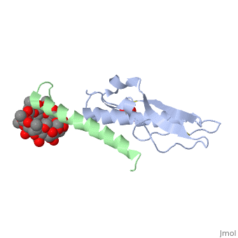
== | ==Crystal structure of the extracellular domain of human GIP receptor in complex with the hormone GIP== | ||
Incretins, endogenous polypeptide hormones released in response to food | <StructureSection load='2qkh' size='340' side='right'caption='[[2qkh]], [[Resolution|resolution]] 1.90Å' scene=''> | ||
== Structural highlights == | |||
<table><tr><td colspan='2'>[[2qkh]] is a 2 chain structure with sequence from [https://en.wikipedia.org/wiki/Homo_sapiens Homo sapiens]. Full crystallographic information is available from [http://oca.weizmann.ac.il/oca-bin/ocashort?id=2QKH OCA]. For a <b>guided tour on the structure components</b> use [https://proteopedia.org/fgij/fg.htm?mol=2QKH FirstGlance]. <br> | |||
</td></tr><tr id='method'><td class="sblockLbl"><b>[[Empirical_models|Method:]]</b></td><td class="sblockDat" id="methodDat">X-ray diffraction, [[Resolution|Resolution]] 1.9Å</td></tr> | |||
<tr id='ligand'><td class="sblockLbl"><b>[[Ligand|Ligands:]]</b></td><td class="sblockDat" id="ligandDat"><scene name='pdbligand=GLC:ALPHA-D-GLUCOSE'>GLC</scene>, <scene name='pdbligand=PRD_900086:methyl-beta-cyclodextrin'>PRD_900086</scene>, <scene name='pdbligand=TAR:D(-)-TARTARIC+ACID'>TAR</scene>, <scene name='pdbligand=ZB0:(2S,3R,4S,5R,6R)-6-(hydroxymethyl)-3,4-dimethoxy-oxane-2,5-diol'>ZB0</scene>, <scene name='pdbligand=ZB1:(2S,3R,4S,5R,6R)-6-(hydroxymethyl)-4-methoxy-oxane-2,3,5-triol'>ZB1</scene>, <scene name='pdbligand=ZB2:2-O-methyl-alpha-D-glucopyranose'>ZB2</scene>, <scene name='pdbligand=ZB3:(2S,3R,4S,5S,6R)-3-methoxy-6-(methoxymethyl)oxane-2,4,5-triol'>ZB3</scene></td></tr> | |||
<tr id='resources'><td class="sblockLbl"><b>Resources:</b></td><td class="sblockDat"><span class='plainlinks'>[https://proteopedia.org/fgij/fg.htm?mol=2qkh FirstGlance], [http://oca.weizmann.ac.il/oca-bin/ocaids?id=2qkh OCA], [https://pdbe.org/2qkh PDBe], [https://www.rcsb.org/pdb/explore.do?structureId=2qkh RCSB], [https://www.ebi.ac.uk/pdbsum/2qkh PDBsum], [https://prosat.h-its.org/prosat/prosatexe?pdbcode=2qkh ProSAT]</span></td></tr> | |||
</table> | |||
== Function == | |||
[https://www.uniprot.org/uniprot/GIP_HUMAN GIP_HUMAN] Potent stimulator of insulin secretion and relatively poor inhibitor of gastric acid secretion. | |||
== Evolutionary Conservation == | |||
[[Image:Consurf_key_small.gif|200px|right]] | |||
Check<jmol> | |||
<jmolCheckbox> | |||
<scriptWhenChecked>; select protein; define ~consurf_to_do selected; consurf_initial_scene = true; script "/wiki/ConSurf/qk/2qkh_consurf.spt"</scriptWhenChecked> | |||
<scriptWhenUnchecked>script /wiki/extensions/Proteopedia/spt/initialview03.spt</scriptWhenUnchecked> | |||
<text>to colour the structure by Evolutionary Conservation</text> | |||
</jmolCheckbox> | |||
</jmol>, as determined by [http://consurfdb.tau.ac.il/ ConSurfDB]. You may read the [[Conservation%2C_Evolutionary|explanation]] of the method and the full data available from [http://bental.tau.ac.il/new_ConSurfDB/main_output.php?pdb_ID=2qkh ConSurf]. | |||
<div style="clear:both"></div> | |||
<div style="background-color:#fffaf0;"> | |||
== Publication Abstract from PubMed == | |||
Incretins, endogenous polypeptide hormones released in response to food intake, potentiate insulin secretion from pancreatic beta cells after oral glucose ingestion (the incretin effect). This response is signaled by the two peptide hormones glucose-dependent insulinotropic polypeptide (GIP) (also known as gastric inhibitory polypeptide) and glucagon-like peptide 1 through binding and activation of their cognate class 2 G protein-coupled receptors (GPCRs). Because the incretin effect is lost or significantly reduced in patients with type 2 diabetes mellitus, glucagon-like peptide 1 and GIP have attracted considerable attention for their potential in antidiabetic therapy. A paucity of structural information precludes a detailed understanding of the processes of hormone binding and receptor activation, hampering efforts to develop novel pharmaceuticals. Here we report the crystal structure of the complex of human GIP receptor extracellular domain (ECD) with its agonist, the incretin GIP(1-42). The hormone binds in an alpha-helical conformation in a surface groove of the ECD largely through hydrophobic interactions. The N-terminal ligand residues would remain free to interact with other parts of the receptor. Thermodynamic data suggest that binding is concomitant with structural organization of the hormone, resulting in a complex mode of receptor-ligand recognition. The presentation of a well structured, alpha-helical ligand by the ECD is expected to be conserved among other hormone receptors of this class. | |||
Crystal structure of the incretin-bound extracellular domain of a G protein-coupled receptor.,Parthier C, Kleinschmidt M, Neumann P, Rudolph R, Manhart S, Schlenzig D, Fanghanel J, Rahfeld JU, Demuth HU, Stubbs MT Proc Natl Acad Sci U S A. 2007 Aug 28;104(35):13942-7. Epub 2007 Aug 21. PMID:17715056<ref>PMID:17715056</ref> | |||
From MEDLINE®/PubMed®, a database of the U.S. National Library of Medicine.<br> | |||
</div> | |||
<div class="pdbe-citations 2qkh" style="background-color:#fffaf0;"></div> | |||
==See Also== | |||
*[[Glucose-dependent Insulinotropic Polypeptide Receptor|Glucose-dependent Insulinotropic Polypeptide Receptor]] | |||
== References == | |||
<references/> | |||
__TOC__ | |||
</StructureSection> | |||
[[Category: Homo sapiens]] | [[Category: Homo sapiens]] | ||
[[Category: | [[Category: Large Structures]] | ||
[[Category: Demuth | [[Category: Demuth H-U]] | ||
[[Category: Fanghanel | [[Category: Fanghanel J]] | ||
[[Category: Kleinschmidt | [[Category: Kleinschmidt M]] | ||
[[Category: Manhart | [[Category: Manhart S]] | ||
[[Category: Neumann | [[Category: Neumann P]] | ||
[[Category: Parthier | [[Category: Parthier C]] | ||
[[Category: Rahfeld | [[Category: Rahfeld J-U]] | ||
[[Category: Rudolph | [[Category: Rudolph R]] | ||
[[Category: Schlenzig | [[Category: Schlenzig D]] | ||
[[Category: Stubbs | [[Category: Stubbs MT]] | ||
Latest revision as of 11:33, 30 October 2024
Crystal structure of the extracellular domain of human GIP receptor in complex with the hormone GIPCrystal structure of the extracellular domain of human GIP receptor in complex with the hormone GIP
Structural highlights
FunctionGIP_HUMAN Potent stimulator of insulin secretion and relatively poor inhibitor of gastric acid secretion. Evolutionary Conservation Check, as determined by ConSurfDB. You may read the explanation of the method and the full data available from ConSurf. Publication Abstract from PubMedIncretins, endogenous polypeptide hormones released in response to food intake, potentiate insulin secretion from pancreatic beta cells after oral glucose ingestion (the incretin effect). This response is signaled by the two peptide hormones glucose-dependent insulinotropic polypeptide (GIP) (also known as gastric inhibitory polypeptide) and glucagon-like peptide 1 through binding and activation of their cognate class 2 G protein-coupled receptors (GPCRs). Because the incretin effect is lost or significantly reduced in patients with type 2 diabetes mellitus, glucagon-like peptide 1 and GIP have attracted considerable attention for their potential in antidiabetic therapy. A paucity of structural information precludes a detailed understanding of the processes of hormone binding and receptor activation, hampering efforts to develop novel pharmaceuticals. Here we report the crystal structure of the complex of human GIP receptor extracellular domain (ECD) with its agonist, the incretin GIP(1-42). The hormone binds in an alpha-helical conformation in a surface groove of the ECD largely through hydrophobic interactions. The N-terminal ligand residues would remain free to interact with other parts of the receptor. Thermodynamic data suggest that binding is concomitant with structural organization of the hormone, resulting in a complex mode of receptor-ligand recognition. The presentation of a well structured, alpha-helical ligand by the ECD is expected to be conserved among other hormone receptors of this class. Crystal structure of the incretin-bound extracellular domain of a G protein-coupled receptor.,Parthier C, Kleinschmidt M, Neumann P, Rudolph R, Manhart S, Schlenzig D, Fanghanel J, Rahfeld JU, Demuth HU, Stubbs MT Proc Natl Acad Sci U S A. 2007 Aug 28;104(35):13942-7. Epub 2007 Aug 21. PMID:17715056[1] From MEDLINE®/PubMed®, a database of the U.S. National Library of Medicine. See AlsoReferences
|
| ||||||||||||||||||