2v9d: Difference between revisions
Jump to navigation
Jump to search
New page: left|200px <!-- The line below this paragraph, containing "STRUCTURE_2v9d", creates the "Structure Box" on the page. You may change the PDB parameter (which sets the PD... |
No edit summary |
||
| (10 intermediate revisions by the same user not shown) | |||
| Line 1: | Line 1: | ||
< | ==Crystal Structure of YagE, a prophage protein belonging to the dihydrodipicolinic acid synthase family from E. coli K12== | ||
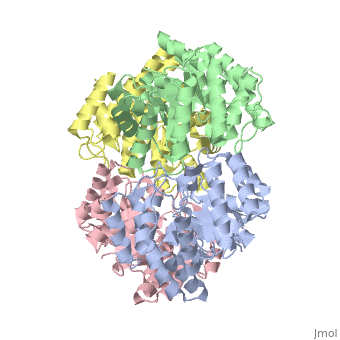
<StructureSection load='2v9d' size='340' side='right'caption='[[2v9d]], [[Resolution|resolution]] 2.15Å' scene=''> | |||
== Structural highlights == | |||
<table><tr><td colspan='2'>[[2v9d]] is a 4 chain structure with sequence from [https://en.wikipedia.org/wiki/Escherichia_coli_K-12 Escherichia coli K-12]. Full crystallographic information is available from [http://oca.weizmann.ac.il/oca-bin/ocashort?id=2V9D OCA]. For a <b>guided tour on the structure components</b> use [https://proteopedia.org/fgij/fg.htm?mol=2V9D FirstGlance]. <br> | |||
</td></tr><tr id='method'><td class="sblockLbl"><b>[[Empirical_models|Method:]]</b></td><td class="sblockDat" id="methodDat">X-ray diffraction, [[Resolution|Resolution]] 2.15Å</td></tr> | |||
<tr id='ligand'><td class="sblockLbl"><b>[[Ligand|Ligands:]]</b></td><td class="sblockDat" id="ligandDat"><scene name='pdbligand=MSE:SELENOMETHIONINE'>MSE</scene></td></tr> | |||
<tr id='resources'><td class="sblockLbl"><b>Resources:</b></td><td class="sblockDat"><span class='plainlinks'>[https://proteopedia.org/fgij/fg.htm?mol=2v9d FirstGlance], [http://oca.weizmann.ac.il/oca-bin/ocaids?id=2v9d OCA], [https://pdbe.org/2v9d PDBe], [https://www.rcsb.org/pdb/explore.do?structureId=2v9d RCSB], [https://www.ebi.ac.uk/pdbsum/2v9d PDBsum], [https://prosat.h-its.org/prosat/prosatexe?pdbcode=2v9d ProSAT]</span></td></tr> | |||
</table> | |||
== Function == | |||
[https://www.uniprot.org/uniprot/YAGE_ECOLI YAGE_ECOLI] Catalyzes the formation of 2-keto-3-deoxy-galactonate (KDGal) from pyruvate and glyceraldehyde. Overexpression leads to increased growth (over 2 hours) in the presence of the antibiotics norfloxacin, ampicillin and streptomycin. | |||
== Evolutionary Conservation == | |||
[[Image:Consurf_key_small.gif|200px|right]] | |||
Check<jmol> | |||
<jmolCheckbox> | |||
<scriptWhenChecked>; select protein; define ~consurf_to_do selected; consurf_initial_scene = true; script "/wiki/ConSurf/v9/2v9d_consurf.spt"</scriptWhenChecked> | |||
<scriptWhenUnchecked>script /wiki/extensions/Proteopedia/spt/initialview03.spt</scriptWhenUnchecked> | |||
<text>to colour the structure by Evolutionary Conservation</text> | |||
</jmolCheckbox> | |||
</jmol>, as determined by [http://consurfdb.tau.ac.il/ ConSurfDB]. You may read the [[Conservation%2C_Evolutionary|explanation]] of the method and the full data available from [http://bental.tau.ac.il/new_ConSurfDB/main_output.php?pdb_ID=2v9d ConSurf]. | |||
<div style="clear:both"></div> | |||
== | ==See Also== | ||
*[[Dihydrodipicolinate synthase|Dihydrodipicolinate synthase]] | |||
__TOC__ | |||
</StructureSection> | |||
[[ | [[Category: Escherichia coli K-12]] | ||
[[Category: Large Structures]] | |||
[[Category: Albeck S]] | |||
< | [[Category: Bourenkov G]] | ||
[[Category: Escherichia coli]] | [[Category: Dym O]] | ||
[[Category: Albeck | [[Category: Greenblatt HM]] | ||
[[Category: Bourenkov | [[Category: Krishnaswamy S]] | ||
[[Category: Dym | [[Category: Lamzin V]] | ||
[[Category: Greenblatt | [[Category: Manicka S]] | ||
[[Category: Krishnaswamy | [[Category: Peleg Y]] | ||
[[Category: Lamzin | [[Category: Sussman JL]] | ||
[[Category: Manicka | [[Category: Unger T]] | ||
[[Category: Peleg | |||
[[Category: Sussman | |||
[[Category: Unger | |||
Latest revision as of 04:26, 21 November 2024
Crystal Structure of YagE, a prophage protein belonging to the dihydrodipicolinic acid synthase family from E. coli K12Crystal Structure of YagE, a prophage protein belonging to the dihydrodipicolinic acid synthase family from E. coli K12
Structural highlights
FunctionYAGE_ECOLI Catalyzes the formation of 2-keto-3-deoxy-galactonate (KDGal) from pyruvate and glyceraldehyde. Overexpression leads to increased growth (over 2 hours) in the presence of the antibiotics norfloxacin, ampicillin and streptomycin. Evolutionary Conservation Check, as determined by ConSurfDB. You may read the explanation of the method and the full data available from ConSurf. See Also |
| ||||||||||||||||||