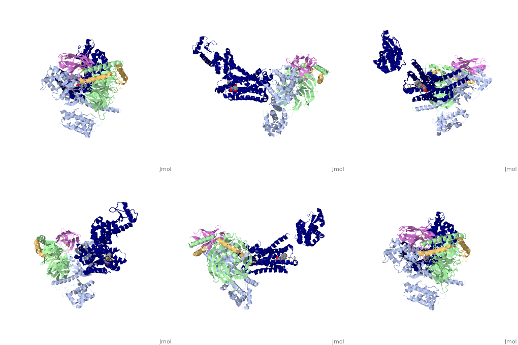
3sn6: Difference between revisions
No edit summary |
No edit summary |
||
| (9 intermediate revisions by the same user not shown) | |||
| Line 1: | Line 1: | ||
==Crystal structure of the beta2 adrenergic receptor-Gs protein complex== | |||
<StructureSection load='3sn6' size='340' side='right'caption='[[3sn6]], [[Resolution|resolution]] 3.20Å' scene=''> | |||
== Structural highlights == | |||
<table><tr><td colspan='2'>[[3sn6]] is a 5 chain structure with sequence from [https://en.wikipedia.org/wiki/Bos_taurus Bos taurus], [https://en.wikipedia.org/wiki/Escherichia_virus_T4 Escherichia virus T4], [https://en.wikipedia.org/wiki/Homo_sapiens Homo sapiens], [https://en.wikipedia.org/wiki/Lama_glama Lama glama] and [https://en.wikipedia.org/wiki/Rattus_norvegicus Rattus norvegicus]. Full crystallographic information is available from [http://oca.weizmann.ac.il/oca-bin/ocashort?id=3SN6 OCA]. For a <b>guided tour on the structure components</b> use [https://proteopedia.org/fgij/fg.htm?mol=3SN6 FirstGlance]. <br> | |||
</td></tr><tr id='method'><td class="sblockLbl"><b>[[Empirical_models|Method:]]</b></td><td class="sblockDat" id="methodDat">X-ray diffraction, [[Resolution|Resolution]] 3.2Å</td></tr> | |||
<tr id='ligand'><td class="sblockLbl"><b>[[Ligand|Ligands:]]</b></td><td class="sblockDat" id="ligandDat"><scene name='pdbligand=P0G:8-[(1R)-2-{[1,1-DIMETHYL-2-(2-METHYLPHENYL)ETHYL]AMINO}-1-HYDROXYETHYL]-5-HYDROXY-2H-1,4-BENZOXAZIN-3(4H)-ONE'>P0G</scene></td></tr> | |||
<tr id='resources'><td class="sblockLbl"><b>Resources:</b></td><td class="sblockDat"><span class='plainlinks'>[https://proteopedia.org/fgij/fg.htm?mol=3sn6 FirstGlance], [http://oca.weizmann.ac.il/oca-bin/ocaids?id=3sn6 OCA], [https://pdbe.org/3sn6 PDBe], [https://www.rcsb.org/pdb/explore.do?structureId=3sn6 RCSB], [https://www.ebi.ac.uk/pdbsum/3sn6 PDBsum], [https://prosat.h-its.org/prosat/prosatexe?pdbcode=3sn6 ProSAT]</span></td></tr> | |||
</table> | |||
== Function == | |||
[https://www.uniprot.org/uniprot/GNAS2_BOVIN GNAS2_BOVIN] Guanine nucleotide-binding proteins (G proteins) are involved as modulators or transducers in various transmembrane signaling systems. The G(s) protein is involved in hormonal regulation of adenylate cyclase: it activates the cyclase in response to beta-adrenergic stimuli. | |||
<div style="background-color:#fffaf0;"> | |||
== Publication Abstract from PubMed == | |||
G protein-coupled receptors (GPCRs) are responsible for the majority of cellular responses to hormones and neurotransmitters as well as the senses of sight, olfaction and taste. The paradigm of GPCR signalling is the activation of a heterotrimeric GTP binding protein (G protein) by an agonist-occupied receptor. The beta(2) adrenergic receptor (beta(2)AR) activation of Gs, the stimulatory G protein for adenylyl cyclase, has long been a model system for GPCR signalling. Here we present the crystal structure of the active state ternary complex composed of agonist-occupied monomeric beta(2)AR and nucleotide-free Gs heterotrimer. The principal interactions between the beta(2)AR and Gs involve the amino- and carboxy-terminal alpha-helices of Gs, with conformational changes propagating to the nucleotide-binding pocket. The largest conformational changes in the beta(2)AR include a 14 A outward movement at the cytoplasmic end of transmembrane segment 6 (TM6) and an alpha-helical extension of the cytoplasmic end of TM5. The most surprising observation is a major displacement of the alpha-helical domain of Galphas relative to the Ras-like GTPase domain. This crystal structure represents the first high-resolution view of transmembrane signalling by a GPCR. | |||
Crystal structure of the beta2 adrenergic receptor-Gs protein complex.,Rasmussen SG, DeVree BT, Zou Y, Kruse AC, Chung KY, Kobilka TS, Thian FS, Chae PS, Pardon E, Calinski D, Mathiesen JM, Shah ST, Lyons JA, Caffrey M, Gellman SH, Steyaert J, Skiniotis G, Weis WI, Sunahara RK, Kobilka BK Nature. 2011 Jul 19;477(7366):549-55. doi: 10.1038/nature10361. PMID:21772288<ref>PMID:21772288</ref> | |||
From MEDLINE®/PubMed®, a database of the U.S. National Library of Medicine.<br> | |||
</div> | |||
<div class="pdbe-citations 3sn6" style="background-color:#fffaf0;"></div> | |||
==See Also== | ==See Also== | ||
*[[Adrenergic receptor|Adrenergic receptor]] | *[[Adrenergic receptor|Adrenergic receptor]] | ||
*[[GTP-binding protein|GTP-binding protein]] | *[[Adrenergic receptor 3D structures|Adrenergic receptor 3D structures]] | ||
*[[ | *[[Antibody 3D structures|Antibody 3D structures]] | ||
*[[ | *[[G protein-coupled receptor|G protein-coupled receptor]] | ||
*[[ | *[[GTP-binding protein 3D structures|GTP-binding protein 3D structures]] | ||
*[[ | *[[Hormone|Hormone]] | ||
*[[Lysozyme 3D structures|Lysozyme 3D structures]] | |||
== | *[[Neurotransmitters|Neurotransmitters]] | ||
< | *[[Transducin 3D structures|Transducin 3D structures]] | ||
*[[3D structures of non-human antibody|3D structures of non-human antibody]] | |||
== References == | |||
<references/> | |||
__TOC__ | |||
</StructureSection> | |||
[[Category: Bos taurus]] | [[Category: Bos taurus]] | ||
[[Category: | [[Category: Escherichia virus T4]] | ||
[[Category: Homo sapiens]] | |||
[[Category: Lama glama]] | [[Category: Lama glama]] | ||
[[Category: | [[Category: Large Structures]] | ||
[[Category: Rattus norvegicus]] | [[Category: Rattus norvegicus]] | ||
[[Category: Caffrey | [[Category: Caffrey M]] | ||
[[Category: Calinski | [[Category: Calinski D]] | ||
[[Category: Chae | [[Category: Chae PS]] | ||
[[Category: Chung | [[Category: Chung KY]] | ||
[[Category: DeVree | [[Category: DeVree BT]] | ||
[[Category: Gellman | [[Category: Gellman SH]] | ||
[[Category: Kobilka | [[Category: Kobilka BK]] | ||
[[Category: Kobilka | [[Category: Kobilka TS]] | ||
[[Category: Kruse | [[Category: Kruse AC]] | ||
[[Category: Lyons | [[Category: Lyons JA]] | ||
[[Category: Mathiesen | [[Category: Mathiesen JM]] | ||
[[Category: Pardon | [[Category: Pardon E]] | ||
[[Category: Rasmussen | [[Category: Rasmussen SGF]] | ||
[[Category: Shah | [[Category: Shah STA]] | ||
[[Category: Skiniotis | [[Category: Skiniotis G]] | ||
[[Category: Steyaert | [[Category: Steyaert J]] | ||
[[Category: Sunahara | [[Category: Sunahara RK]] | ||
[[Category: Thian | [[Category: Thian FS]] | ||
[[Category: Weis | [[Category: Weis WI]] | ||
[[Category: Zou | [[Category: Zou Y]] | ||
Latest revision as of 05:24, 21 November 2024
Crystal structure of the beta2 adrenergic receptor-Gs protein complexCrystal structure of the beta2 adrenergic receptor-Gs protein complex
Structural highlights
FunctionGNAS2_BOVIN Guanine nucleotide-binding proteins (G proteins) are involved as modulators or transducers in various transmembrane signaling systems. The G(s) protein is involved in hormonal regulation of adenylate cyclase: it activates the cyclase in response to beta-adrenergic stimuli. Publication Abstract from PubMedG protein-coupled receptors (GPCRs) are responsible for the majority of cellular responses to hormones and neurotransmitters as well as the senses of sight, olfaction and taste. The paradigm of GPCR signalling is the activation of a heterotrimeric GTP binding protein (G protein) by an agonist-occupied receptor. The beta(2) adrenergic receptor (beta(2)AR) activation of Gs, the stimulatory G protein for adenylyl cyclase, has long been a model system for GPCR signalling. Here we present the crystal structure of the active state ternary complex composed of agonist-occupied monomeric beta(2)AR and nucleotide-free Gs heterotrimer. The principal interactions between the beta(2)AR and Gs involve the amino- and carboxy-terminal alpha-helices of Gs, with conformational changes propagating to the nucleotide-binding pocket. The largest conformational changes in the beta(2)AR include a 14 A outward movement at the cytoplasmic end of transmembrane segment 6 (TM6) and an alpha-helical extension of the cytoplasmic end of TM5. The most surprising observation is a major displacement of the alpha-helical domain of Galphas relative to the Ras-like GTPase domain. This crystal structure represents the first high-resolution view of transmembrane signalling by a GPCR. Crystal structure of the beta2 adrenergic receptor-Gs protein complex.,Rasmussen SG, DeVree BT, Zou Y, Kruse AC, Chung KY, Kobilka TS, Thian FS, Chae PS, Pardon E, Calinski D, Mathiesen JM, Shah ST, Lyons JA, Caffrey M, Gellman SH, Steyaert J, Skiniotis G, Weis WI, Sunahara RK, Kobilka BK Nature. 2011 Jul 19;477(7366):549-55. doi: 10.1038/nature10361. PMID:21772288[1] From MEDLINE®/PubMed®, a database of the U.S. National Library of Medicine. See Also
References
|
| ||||||||||||||||||
