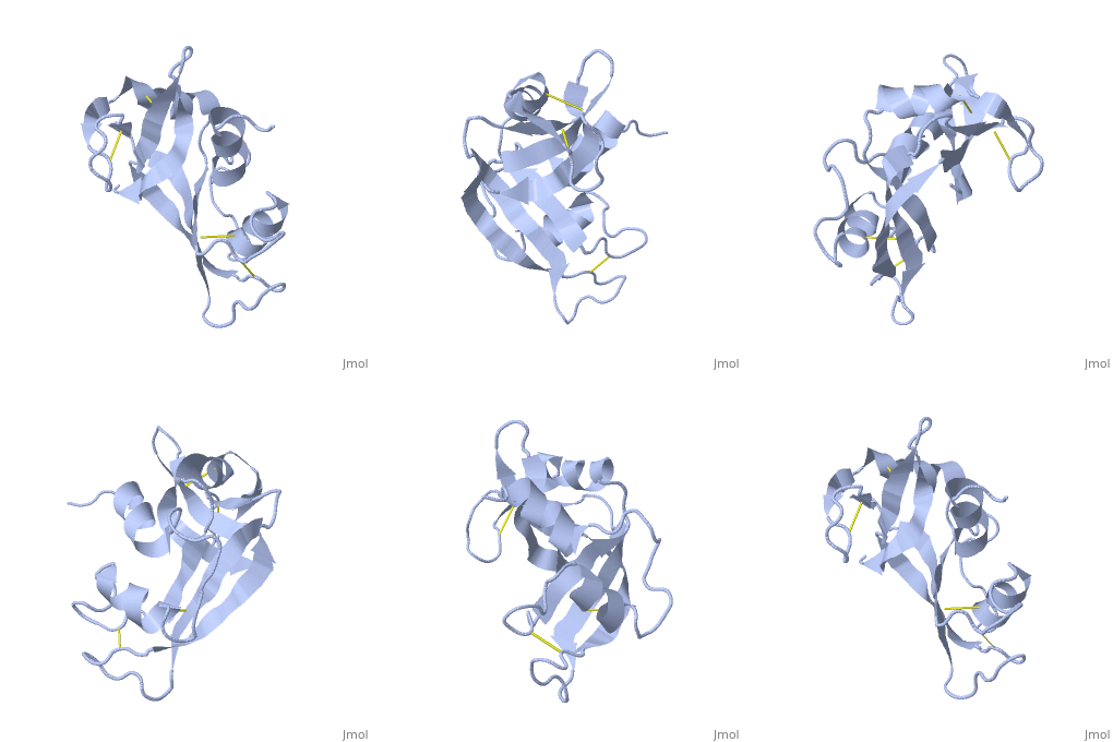
3rat: Difference between revisions
No edit summary |
No edit summary |
||
| (3 intermediate revisions by the same user not shown) | |||
| Line 1: | Line 1: | ||
==EFFECTS OF TEMPERATURE ON PROTEIN STRUCTURE AND DYNAMICS: X-RAY CRYSTALLOGRAPHIC STUDIES OF THE PROTEIN RIBONUCLEASE-A AT NINE DIFFERENT TEMPERATURES FROM 98 TO 320 K== | ==EFFECTS OF TEMPERATURE ON PROTEIN STRUCTURE AND DYNAMICS: X-RAY CRYSTALLOGRAPHIC STUDIES OF THE PROTEIN RIBONUCLEASE-A AT NINE DIFFERENT TEMPERATURES FROM 98 TO 320 K== | ||
<StructureSection load='3rat' size='340' side='right' caption='[[3rat]], [[Resolution|resolution]] 1.50Å' scene=''> | <StructureSection load='3rat' size='340' side='right'caption='[[3rat]], [[Resolution|resolution]] 1.50Å' scene=''> | ||
== Structural highlights == | == Structural highlights == | ||
<table><tr><td colspan='2'>[[3rat]] is a 1 chain structure with sequence from [ | <table><tr><td colspan='2'>[[3rat]] is a 1 chain structure with sequence from [https://en.wikipedia.org/wiki/Bos_taurus Bos taurus]. Full crystallographic information is available from [http://oca.weizmann.ac.il/oca-bin/ocashort?id=3RAT OCA]. For a <b>guided tour on the structure components</b> use [https://proteopedia.org/fgij/fg.htm?mol=3RAT FirstGlance]. <br> | ||
</td></tr><tr id=' | </td></tr><tr id='method'><td class="sblockLbl"><b>[[Empirical_models|Method:]]</b></td><td class="sblockDat" id="methodDat">X-ray diffraction, [[Resolution|Resolution]] 1.5Å</td></tr> | ||
<tr id='resources'><td class="sblockLbl"><b>Resources:</b></td><td class="sblockDat"><span class='plainlinks'>[ | <tr id='resources'><td class="sblockLbl"><b>Resources:</b></td><td class="sblockDat"><span class='plainlinks'>[https://proteopedia.org/fgij/fg.htm?mol=3rat FirstGlance], [http://oca.weizmann.ac.il/oca-bin/ocaids?id=3rat OCA], [https://pdbe.org/3rat PDBe], [https://www.rcsb.org/pdb/explore.do?structureId=3rat RCSB], [https://www.ebi.ac.uk/pdbsum/3rat PDBsum], [https://prosat.h-its.org/prosat/prosatexe?pdbcode=3rat ProSAT]</span></td></tr> | ||
</table> | </table> | ||
== Function == | == Function == | ||
[ | [https://www.uniprot.org/uniprot/RNAS1_BOVIN RNAS1_BOVIN] Endonuclease that catalyzes the cleavage of RNA on the 3' side of pyrimidine nucleotides. Acts on single stranded and double stranded RNA.<ref>PMID:7479688</ref> | ||
== Evolutionary Conservation == | == Evolutionary Conservation == | ||
[[Image:Consurf_key_small.gif|200px|right]] | [[Image:Consurf_key_small.gif|200px|right]] | ||
Check<jmol> | Check<jmol> | ||
<jmolCheckbox> | <jmolCheckbox> | ||
<scriptWhenChecked>select protein; define ~consurf_to_do selected; consurf_initial_scene = true; script "/wiki/ConSurf/ra/3rat_consurf.spt"</scriptWhenChecked> | <scriptWhenChecked>; select protein; define ~consurf_to_do selected; consurf_initial_scene = true; script "/wiki/ConSurf/ra/3rat_consurf.spt"</scriptWhenChecked> | ||
<scriptWhenUnchecked>script /wiki/extensions/Proteopedia/spt/ | <scriptWhenUnchecked>script /wiki/extensions/Proteopedia/spt/initialview03.spt</scriptWhenUnchecked> | ||
<text>to colour the structure by Evolutionary Conservation</text> | <text>to colour the structure by Evolutionary Conservation</text> | ||
</jmolCheckbox> | </jmolCheckbox> | ||
</jmol>, as determined by [http://consurfdb.tau.ac.il/ ConSurfDB]. You may read the [[Conservation%2C_Evolutionary|explanation]] of the method and the full data available from [http://bental.tau.ac.il/new_ConSurfDB/ | </jmol>, as determined by [http://consurfdb.tau.ac.il/ ConSurfDB]. You may read the [[Conservation%2C_Evolutionary|explanation]] of the method and the full data available from [http://bental.tau.ac.il/new_ConSurfDB/main_output.php?pdb_ID=3rat ConSurf]. | ||
<div style="clear:both"></div> | <div style="clear:both"></div> | ||
<div style="background-color:#fffaf0;"> | <div style="background-color:#fffaf0;"> | ||
| Line 26: | Line 27: | ||
From MEDLINE®/PubMed®, a database of the U.S. National Library of Medicine.<br> | From MEDLINE®/PubMed®, a database of the U.S. National Library of Medicine.<br> | ||
</div> | </div> | ||
<div class="pdbe-citations 3rat" style="background-color:#fffaf0;"></div> | |||
==See Also== | ==See Also== | ||
*[[Ribonuclease|Ribonuclease | *[[Ribonuclease 3D structures|Ribonuclease 3D structures]] | ||
== References == | == References == | ||
<references/> | <references/> | ||
| Line 36: | Line 36: | ||
</StructureSection> | </StructureSection> | ||
[[Category: Bos taurus]] | [[Category: Bos taurus]] | ||
[[Category: | [[Category: Large Structures]] | ||
[[Category: Dewan | [[Category: Dewan JC]] | ||
[[Category: Petsko | [[Category: Petsko GA]] | ||
[[Category: Tiltonjunior | [[Category: Tiltonjunior RF]] | ||
Latest revision as of 13:23, 6 November 2024
EFFECTS OF TEMPERATURE ON PROTEIN STRUCTURE AND DYNAMICS: X-RAY CRYSTALLOGRAPHIC STUDIES OF THE PROTEIN RIBONUCLEASE-A AT NINE DIFFERENT TEMPERATURES FROM 98 TO 320 KEFFECTS OF TEMPERATURE ON PROTEIN STRUCTURE AND DYNAMICS: X-RAY CRYSTALLOGRAPHIC STUDIES OF THE PROTEIN RIBONUCLEASE-A AT NINE DIFFERENT TEMPERATURES FROM 98 TO 320 K
Structural highlights
FunctionRNAS1_BOVIN Endonuclease that catalyzes the cleavage of RNA on the 3' side of pyrimidine nucleotides. Acts on single stranded and double stranded RNA.[1] Evolutionary Conservation Check, as determined by ConSurfDB. You may read the explanation of the method and the full data available from ConSurf. Publication Abstract from PubMedStructures using X-ray diffraction data collected to 1.5-A resolution have been determined for the protein ribonuclease-A at nine different temperatures ranging from 98 to 320 K. It is determined that the protein molecule expands slightly (0.4% per 100 K) with increasing temperature and that this expansion is linear. The expansion is due primarily to subtle repacking of the molecule, with exposed and mobile loop regions exhibiting the largest movements. Individual atomic Debye-Waller factors exhibit predominantly biphasic behavior, with a small positive slope at low temperatures and a larger positive slope at higher temperatures. The break in this curve occurs at a characteristic temperature of 180-200 K, perhaps indicative of fundamental changes in the dynamical structure of the surrounding protein solvent. The distribution of protein Debye-Waller factors is observed to broaden as well as shift to higher values as the temperature is increased. Effects of temperature on protein structure and dynamics: X-ray crystallographic studies of the protein ribonuclease-A at nine different temperatures from 98 to 320 K.,Tilton RF Jr, Dewan JC, Petsko GA Biochemistry. 1992 Mar 10;31(9):2469-81. PMID:1547232[2] From MEDLINE®/PubMed®, a database of the U.S. National Library of Medicine. See AlsoReferences
|
| ||||||||||||||||
