1j8s: Difference between revisions
No edit summary |
No edit summary |
||
| (2 intermediate revisions by the same user not shown) | |||
| Line 3: | Line 3: | ||
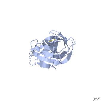
<StructureSection load='1j8s' size='340' side='right'caption='[[1j8s]], [[Resolution|resolution]] 2.10Å' scene=''> | <StructureSection load='1j8s' size='340' side='right'caption='[[1j8s]], [[Resolution|resolution]] 2.10Å' scene=''> | ||
== Structural highlights == | == Structural highlights == | ||
<table><tr><td colspan='2'>[[1j8s]] is a 1 chain structure with sequence from [ | <table><tr><td colspan='2'>[[1j8s]] is a 1 chain structure with sequence from [https://en.wikipedia.org/wiki/Escherichia_coli Escherichia coli]. Full crystallographic information is available from [http://oca.weizmann.ac.il/oca-bin/ocashort?id=1J8S OCA]. For a <b>guided tour on the structure components</b> use [https://proteopedia.org/fgij/fg.htm?mol=1J8S FirstGlance]. <br> | ||
</td></tr><tr id=' | </td></tr><tr id='method'><td class="sblockLbl"><b>[[Empirical_models|Method:]]</b></td><td class="sblockDat" id="methodDat">X-ray diffraction, [[Resolution|Resolution]] 2.1Å</td></tr> | ||
<tr id='resources'><td class="sblockLbl"><b>Resources:</b></td><td class="sblockDat"><span class='plainlinks'>[ | <tr id='ligand'><td class="sblockLbl"><b>[[Ligand|Ligands:]]</b></td><td class="sblockDat" id="ligandDat"><scene name='pdbligand=MSE:SELENOMETHIONINE'>MSE</scene></td></tr> | ||
<tr id='resources'><td class="sblockLbl"><b>Resources:</b></td><td class="sblockDat"><span class='plainlinks'>[https://proteopedia.org/fgij/fg.htm?mol=1j8s FirstGlance], [http://oca.weizmann.ac.il/oca-bin/ocaids?id=1j8s OCA], [https://pdbe.org/1j8s PDBe], [https://www.rcsb.org/pdb/explore.do?structureId=1j8s RCSB], [https://www.ebi.ac.uk/pdbsum/1j8s PDBsum], [https://prosat.h-its.org/prosat/prosatexe?pdbcode=1j8s ProSAT]</span></td></tr> | |||
</table> | </table> | ||
== Function == | |||
[https://www.uniprot.org/uniprot/PAPG2_ECOLX PAPG2_ECOLX] Tip adhesin component of type P pili that plays a critical role in kidney infection through targeted interaction with the globoseries glycolipids containing the Gal-alpha(1-4)-Gal disaccharide present on uroepithelial cells (PubMed:11440716, PubMed:31361021). In turn, transcriptionally regulates host gene expression in kidney cells, leading to inflammatory pathway activation and renal tissue damage. Acts thereby as key determinant of invasive uropathogenic E.coli (UPEC), which cause pyelonephritis and urinary-source bacteremia (By similarity).[UniProtKB:A0A0H2VAQ6]<ref>PMID:11440716</ref> <ref>PMID:31361021</ref> | |||
== Evolutionary Conservation == | == Evolutionary Conservation == | ||
[[Image:Consurf_key_small.gif|200px|right]] | [[Image:Consurf_key_small.gif|200px|right]] | ||
| Line 12: | Line 15: | ||
<jmolCheckbox> | <jmolCheckbox> | ||
<scriptWhenChecked>; select protein; define ~consurf_to_do selected; consurf_initial_scene = true; script "/wiki/ConSurf/j8/1j8s_consurf.spt"</scriptWhenChecked> | <scriptWhenChecked>; select protein; define ~consurf_to_do selected; consurf_initial_scene = true; script "/wiki/ConSurf/j8/1j8s_consurf.spt"</scriptWhenChecked> | ||
<scriptWhenUnchecked>script /wiki/extensions/Proteopedia/spt/ | <scriptWhenUnchecked>script /wiki/extensions/Proteopedia/spt/initialview03.spt</scriptWhenUnchecked> | ||
<text>to colour the structure by Evolutionary Conservation</text> | <text>to colour the structure by Evolutionary Conservation</text> | ||
</jmolCheckbox> | </jmolCheckbox> | ||
| Line 26: | Line 29: | ||
</div> | </div> | ||
<div class="pdbe-citations 1j8s" style="background-color:#fffaf0;"></div> | <div class="pdbe-citations 1j8s" style="background-color:#fffaf0;"></div> | ||
== References == | == References == | ||
<references/> | <references/> | ||
__TOC__ | __TOC__ | ||
</StructureSection> | </StructureSection> | ||
[[Category: | [[Category: Escherichia coli]] | ||
[[Category: Large Structures]] | [[Category: Large Structures]] | ||
[[Category: Dodson | [[Category: Dodson KW]] | ||
[[Category: Hultgren | [[Category: Hultgren SJ]] | ||
[[Category: Magnusson | [[Category: Magnusson G]] | ||
[[Category: Pinkner | [[Category: Pinkner JS]] | ||
[[Category: Rose | [[Category: Rose T]] | ||
[[Category: Waksman | [[Category: Waksman G]] | ||
Latest revision as of 07:38, 17 October 2024
PAPG ADHESIN RECEPTOR BINDING DOMAIN-UNBOUND FORMPAPG ADHESIN RECEPTOR BINDING DOMAIN-UNBOUND FORM
Structural highlights
FunctionPAPG2_ECOLX Tip adhesin component of type P pili that plays a critical role in kidney infection through targeted interaction with the globoseries glycolipids containing the Gal-alpha(1-4)-Gal disaccharide present on uroepithelial cells (PubMed:11440716, PubMed:31361021). In turn, transcriptionally regulates host gene expression in kidney cells, leading to inflammatory pathway activation and renal tissue damage. Acts thereby as key determinant of invasive uropathogenic E.coli (UPEC), which cause pyelonephritis and urinary-source bacteremia (By similarity).[UniProtKB:A0A0H2VAQ6][1] [2] Evolutionary Conservation Check, as determined by ConSurfDB. You may read the explanation of the method and the full data available from ConSurf. Publication Abstract from PubMedPapG is the adhesin at the tip of the P pilus that mediates attachment of uropathogenic Escherichia coli to the uroepithelium of the human kidney. The human specific allele of PapG binds to globoside (GbO4), which consists of the tetrasaccharide GalNAc beta 1-3Gal alpha 1-4Gal beta 1-4Glc linked to ceramide. Here, we present the crystal structures of a binary complex of the PapG receptor binding domain bound to GbO4 as well as the unbound form of the adhesin. The biological importance of each of the residues involved in binding was investigated by site-directed mutagenesis. These studies provide a molecular snapshot of a host-pathogen interaction that determines the tropism of uropathogenic E. coli for the human kidney and is critical to the pathogenesis of pyelonephritis. Structural basis of the interaction of the pyelonephritic E. coli adhesin to its human kidney receptor.,Dodson KW, Pinkner JS, Rose T, Magnusson G, Hultgren SJ, Waksman G Cell. 2001 Jun 15;105(6):733-43. PMID:11440716[3] From MEDLINE®/PubMed®, a database of the U.S. National Library of Medicine. References
|
| ||||||||||||||||||