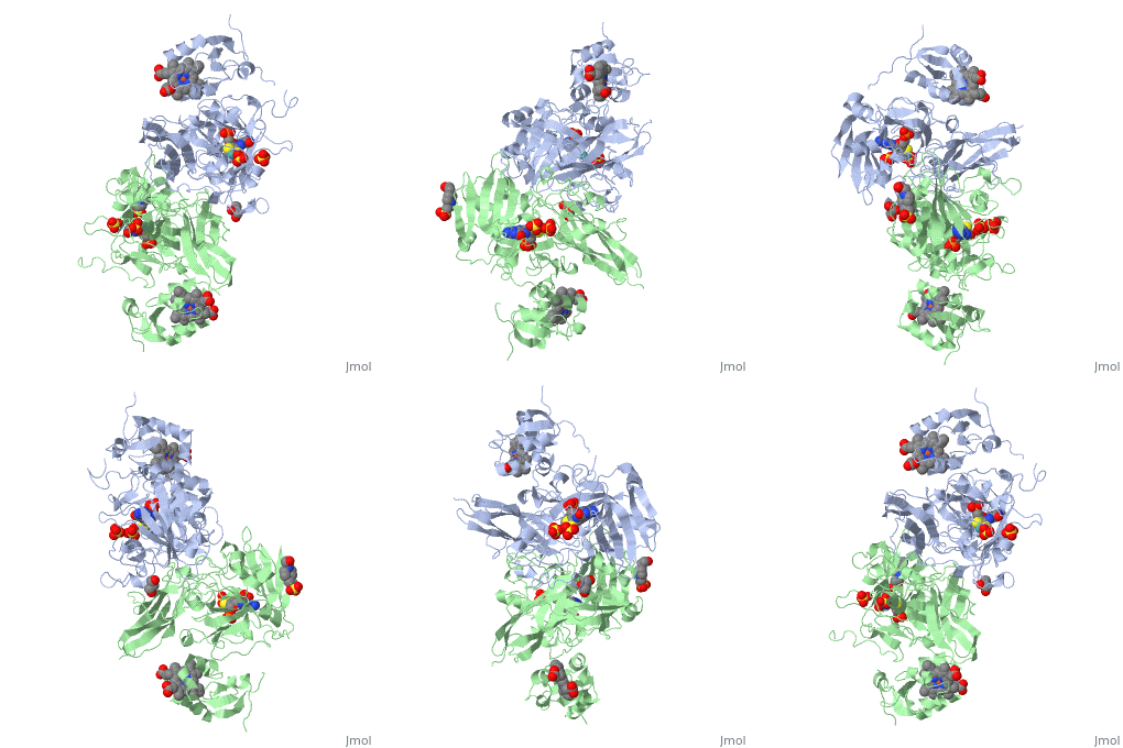
1sox: Difference between revisions
No edit summary |
No edit summary |
||
| (7 intermediate revisions by the same user not shown) | |||
| Line 1: | Line 1: | ||
==SULFITE OXIDASE FROM CHICKEN LIVER== | ==SULFITE OXIDASE FROM CHICKEN LIVER== | ||
<StructureSection load='1sox' size='340' side='right' caption='[[1sox]], [[Resolution|resolution]] 1.90Å' scene=''> | <StructureSection load='1sox' size='340' side='right'caption='[[1sox]], [[Resolution|resolution]] 1.90Å' scene=''> | ||
== Structural highlights == | == Structural highlights == | ||
<table><tr><td colspan='2'>[[1sox]] is a 2 chain structure with sequence from [ | <table><tr><td colspan='2'>[[1sox]] is a 2 chain structure with sequence from [https://en.wikipedia.org/wiki/Gallus_gallus Gallus gallus]. Full crystallographic information is available from [http://oca.weizmann.ac.il/oca-bin/ocashort?id=1SOX OCA]. For a <b>guided tour on the structure components</b> use [https://proteopedia.org/fgij/fg.htm?mol=1SOX FirstGlance]. <br> | ||
</td></tr><tr><td class="sblockLbl"><b>[[Ligand|Ligands:]]</b></td><td class="sblockDat"><scene name='pdbligand=EPE:4-(2-HYDROXYETHYL)-1-PIPERAZINE+ETHANESULFONIC+ACID'>EPE</scene>, <scene name='pdbligand=GOL:GLYCEROL'>GOL</scene>, <scene name='pdbligand=HEM:PROTOPORPHYRIN+IX+CONTAINING+FE'>HEM</scene>, <scene name='pdbligand=MO:MOLYBDENUM+ATOM'>MO</scene>, <scene name='pdbligand=MTE:PHOSPHONIC+ACIDMONO-(2-AMINO-5,6-DIMERCAPTO-4-OXO-3,7,8A,9,10,10A-HEXAHYDRO-4H-8-OXA-1,3,9,10-TETRAAZA-ANTHRACEN-7-YLMETHYL)ESTER'>MTE</scene>, <scene name='pdbligand=SO4:SULFATE+ION'>SO4</scene | </td></tr><tr id='method'><td class="sblockLbl"><b>[[Empirical_models|Method:]]</b></td><td class="sblockDat" id="methodDat">X-ray diffraction, [[Resolution|Resolution]] 1.9Å</td></tr> | ||
<tr id='ligand'><td class="sblockLbl"><b>[[Ligand|Ligands:]]</b></td><td class="sblockDat" id="ligandDat"><scene name='pdbligand=EPE:4-(2-HYDROXYETHYL)-1-PIPERAZINE+ETHANESULFONIC+ACID'>EPE</scene>, <scene name='pdbligand=GOL:GLYCEROL'>GOL</scene>, <scene name='pdbligand=HEM:PROTOPORPHYRIN+IX+CONTAINING+FE'>HEM</scene>, <scene name='pdbligand=MO:MOLYBDENUM+ATOM'>MO</scene>, <scene name='pdbligand=MTE:PHOSPHONIC+ACIDMONO-(2-AMINO-5,6-DIMERCAPTO-4-OXO-3,7,8A,9,10,10A-HEXAHYDRO-4H-8-OXA-1,3,9,10-TETRAAZA-ANTHRACEN-7-YLMETHYL)ESTER'>MTE</scene>, <scene name='pdbligand=SO4:SULFATE+ION'>SO4</scene></td></tr> | |||
<tr><td class="sblockLbl"><b>Resources:</b></td><td class="sblockDat"><span class='plainlinks'>[ | <tr id='resources'><td class="sblockLbl"><b>Resources:</b></td><td class="sblockDat"><span class='plainlinks'>[https://proteopedia.org/fgij/fg.htm?mol=1sox FirstGlance], [http://oca.weizmann.ac.il/oca-bin/ocaids?id=1sox OCA], [https://pdbe.org/1sox PDBe], [https://www.rcsb.org/pdb/explore.do?structureId=1sox RCSB], [https://www.ebi.ac.uk/pdbsum/1sox PDBsum], [https://prosat.h-its.org/prosat/prosatexe?pdbcode=1sox ProSAT]</span></td></tr> | ||
<table> | </table> | ||
== Function == | |||
[https://www.uniprot.org/uniprot/SUOX_CHICK SUOX_CHICK] | |||
== Evolutionary Conservation == | == Evolutionary Conservation == | ||
[[Image:Consurf_key_small.gif|200px|right]] | [[Image:Consurf_key_small.gif|200px|right]] | ||
Check<jmol> | Check<jmol> | ||
<jmolCheckbox> | <jmolCheckbox> | ||
<scriptWhenChecked>select protein; define ~consurf_to_do selected; consurf_initial_scene = true; script "/wiki/ConSurf/so/1sox_consurf.spt"</scriptWhenChecked> | <scriptWhenChecked>; select protein; define ~consurf_to_do selected; consurf_initial_scene = true; script "/wiki/ConSurf/so/1sox_consurf.spt"</scriptWhenChecked> | ||
<scriptWhenUnchecked>script /wiki/extensions/Proteopedia/spt/initialview01.spt</scriptWhenUnchecked> | <scriptWhenUnchecked>script /wiki/extensions/Proteopedia/spt/initialview01.spt</scriptWhenUnchecked> | ||
<text>to colour the structure by Evolutionary Conservation</text> | <text>to colour the structure by Evolutionary Conservation</text> | ||
</jmolCheckbox> | </jmolCheckbox> | ||
</jmol>, as determined by [http://consurfdb.tau.ac.il/ ConSurfDB]. You may read the [[Conservation%2C_Evolutionary|explanation]] of the method and the full data available from [http://bental.tau.ac.il/new_ConSurfDB/ | </jmol>, as determined by [http://consurfdb.tau.ac.il/ ConSurfDB]. You may read the [[Conservation%2C_Evolutionary|explanation]] of the method and the full data available from [http://bental.tau.ac.il/new_ConSurfDB/main_output.php?pdb_ID=1sox ConSurf]. | ||
<div style="clear:both"></div> | <div style="clear:both"></div> | ||
<div style="background-color:#fffaf0;"> | <div style="background-color:#fffaf0;"> | ||
| Line 25: | Line 28: | ||
From MEDLINE®/PubMed®, a database of the U.S. National Library of Medicine.<br> | From MEDLINE®/PubMed®, a database of the U.S. National Library of Medicine.<br> | ||
</div> | </div> | ||
<div class="pdbe-citations 1sox" style="background-color:#fffaf0;"></div> | |||
==See Also== | ==See Also== | ||
| Line 33: | Line 37: | ||
</StructureSection> | </StructureSection> | ||
[[Category: Gallus gallus]] | [[Category: Gallus gallus]] | ||
[[Category: | [[Category: Large Structures]] | ||
[[Category: Kisker | [[Category: Kisker C]] | ||
[[Category: Rees | [[Category: Rees DC]] | ||
[[Category: Schindelin | [[Category: Schindelin H]] | ||
Latest revision as of 12:09, 22 May 2024
SULFITE OXIDASE FROM CHICKEN LIVERSULFITE OXIDASE FROM CHICKEN LIVER
Structural highlights
FunctionEvolutionary Conservation Check, as determined by ConSurfDB. You may read the explanation of the method and the full data available from ConSurf. Publication Abstract from PubMedThe molybdenum-containing enzyme sulfite oxidase catalyzes the conversion of sulfite to sulfate, the terminal step in the oxidative degradation of cysteine and methionine. Deficiency of this enzyme in humans usually leads to major neurological abnormalities and early death. The crystal structure of chicken liver sulfite oxidase at 1.9 A resolution reveals that each monomer of the dimeric enzyme consists of three domains. At the active site, the Mo is penta-coordinated by three sulfur ligands, one oxo group, and one water/hydroxo. A sulfate molecule adjacent to the Mo identifies the substrate binding pocket. Four variants associated with sulfite oxidase deficiency have been identified: two mutations are near the sulfate binding site, while the other mutations occur within the domain mediating dimerization. Molecular basis of sulfite oxidase deficiency from the structure of sulfite oxidase.,Kisker C, Schindelin H, Pacheco A, Wehbi WA, Garrett RM, Rajagopalan KV, Enemark JH, Rees DC Cell. 1997 Dec 26;91(7):973-83. PMID:9428520[1] From MEDLINE®/PubMed®, a database of the U.S. National Library of Medicine. See AlsoReferences |
| ||||||||||||||||||
