5dfr: Difference between revisions
Jump to navigation
Jump to search
No edit summary |
No edit summary |
||
| (One intermediate revision by the same user not shown) | |||
| Line 1: | Line 1: | ||
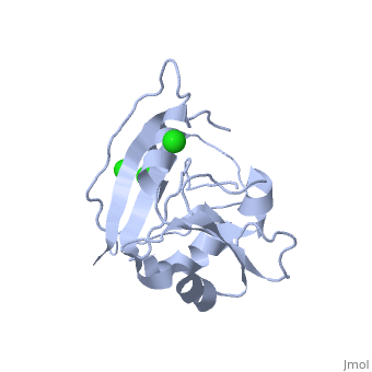
==CRYSTAL STRUCTURE OF UNLIGANDED ESCHERICHIA COLI DIHYDROFOLATE REDUCTASE. LIGAND-INDUCED CONFORMATIONAL CHANGES AND COOPERATIVITY IN BINDING== | ==CRYSTAL STRUCTURE OF UNLIGANDED ESCHERICHIA COLI DIHYDROFOLATE REDUCTASE. LIGAND-INDUCED CONFORMATIONAL CHANGES AND COOPERATIVITY IN BINDING== | ||
<StructureSection load='5dfr' size='340' side='right' caption='[[5dfr]], [[Resolution|resolution]] 2.30Å' scene=''> | <StructureSection load='5dfr' size='340' side='right'caption='[[5dfr]], [[Resolution|resolution]] 2.30Å' scene=''> | ||
== Structural highlights == | == Structural highlights == | ||
<table><tr><td colspan='2'>[[5dfr]] is a 1 chain structure with sequence from [ | <table><tr><td colspan='2'>[[5dfr]] is a 1 chain structure with sequence from [https://en.wikipedia.org/wiki/Escherichia_coli Escherichia coli]. Full crystallographic information is available from [http://oca.weizmann.ac.il/oca-bin/ocashort?id=5DFR OCA]. For a <b>guided tour on the structure components</b> use [https://proteopedia.org/fgij/fg.htm?mol=5DFR FirstGlance]. <br> | ||
</td></tr><tr id=' | </td></tr><tr id='method'><td class="sblockLbl"><b>[[Empirical_models|Method:]]</b></td><td class="sblockDat" id="methodDat">X-ray diffraction, [[Resolution|Resolution]] 2.3Å</td></tr> | ||
<tr id=' | <tr id='ligand'><td class="sblockLbl"><b>[[Ligand|Ligands:]]</b></td><td class="sblockDat" id="ligandDat"><scene name='pdbligand=CL:CHLORIDE+ION'>CL</scene></td></tr> | ||
<tr id='resources'><td class="sblockLbl"><b>Resources:</b></td><td class="sblockDat"><span class='plainlinks'>[ | <tr id='resources'><td class="sblockLbl"><b>Resources:</b></td><td class="sblockDat"><span class='plainlinks'>[https://proteopedia.org/fgij/fg.htm?mol=5dfr FirstGlance], [http://oca.weizmann.ac.il/oca-bin/ocaids?id=5dfr OCA], [https://pdbe.org/5dfr PDBe], [https://www.rcsb.org/pdb/explore.do?structureId=5dfr RCSB], [https://www.ebi.ac.uk/pdbsum/5dfr PDBsum], [https://prosat.h-its.org/prosat/prosatexe?pdbcode=5dfr ProSAT]</span></td></tr> | ||
</table> | </table> | ||
== Function == | == Function == | ||
[ | [https://www.uniprot.org/uniprot/DYR_ECOLI DYR_ECOLI] Key enzyme in folate metabolism. Catalyzes an essential reaction for de novo glycine and purine synthesis, and for DNA precursor synthesis. | ||
== Evolutionary Conservation == | == Evolutionary Conservation == | ||
[[Image:Consurf_key_small.gif|200px|right]] | [[Image:Consurf_key_small.gif|200px|right]] | ||
Check<jmol> | Check<jmol> | ||
<jmolCheckbox> | <jmolCheckbox> | ||
<scriptWhenChecked>select protein; define ~consurf_to_do selected; consurf_initial_scene = true; script "/wiki/ConSurf/df/5dfr_consurf.spt"</scriptWhenChecked> | <scriptWhenChecked>; select protein; define ~consurf_to_do selected; consurf_initial_scene = true; script "/wiki/ConSurf/df/5dfr_consurf.spt"</scriptWhenChecked> | ||
<scriptWhenUnchecked>script /wiki/extensions/Proteopedia/spt/initialview01.spt</scriptWhenUnchecked> | <scriptWhenUnchecked>script /wiki/extensions/Proteopedia/spt/initialview01.spt</scriptWhenUnchecked> | ||
<text>to colour the structure by Evolutionary Conservation</text> | <text>to colour the structure by Evolutionary Conservation</text> | ||
| Line 20: | Line 20: | ||
</jmol>, as determined by [http://consurfdb.tau.ac.il/ ConSurfDB]. You may read the [[Conservation%2C_Evolutionary|explanation]] of the method and the full data available from [http://bental.tau.ac.il/new_ConSurfDB/main_output.php?pdb_ID=5dfr ConSurf]. | </jmol>, as determined by [http://consurfdb.tau.ac.il/ ConSurfDB]. You may read the [[Conservation%2C_Evolutionary|explanation]] of the method and the full data available from [http://bental.tau.ac.il/new_ConSurfDB/main_output.php?pdb_ID=5dfr ConSurf]. | ||
<div style="clear:both"></div> | <div style="clear:both"></div> | ||
==See Also== | ==See Also== | ||
*[[ | *[[Dihydrofolate reductase 3D structures|Dihydrofolate reductase 3D structures]] | ||
__TOC__ | __TOC__ | ||
</StructureSection> | </StructureSection> | ||
[[Category: | [[Category: Escherichia coli]] | ||
[[Category: | [[Category: Large Structures]] | ||
[[Category: Bystroff | [[Category: Bystroff C]] | ||
[[Category: Kraut | [[Category: Kraut J]] | ||
Latest revision as of 15:24, 6 March 2024
CRYSTAL STRUCTURE OF UNLIGANDED ESCHERICHIA COLI DIHYDROFOLATE REDUCTASE. LIGAND-INDUCED CONFORMATIONAL CHANGES AND COOPERATIVITY IN BINDINGCRYSTAL STRUCTURE OF UNLIGANDED ESCHERICHIA COLI DIHYDROFOLATE REDUCTASE. LIGAND-INDUCED CONFORMATIONAL CHANGES AND COOPERATIVITY IN BINDING
Structural highlights
FunctionDYR_ECOLI Key enzyme in folate metabolism. Catalyzes an essential reaction for de novo glycine and purine synthesis, and for DNA precursor synthesis. Evolutionary Conservation Check, as determined by ConSurfDB. You may read the explanation of the method and the full data available from ConSurf. See Also |
| ||||||||||||||||||