2fom: Difference between revisions
Jump to navigation
Jump to search
No edit summary |
No edit summary |
||
| (6 intermediate revisions by the same user not shown) | |||
| Line 1: | Line 1: | ||
==Dengue Virus NS2B/NS3 Protease== | ==Dengue Virus NS2B/NS3 Protease== | ||
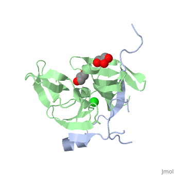
<StructureSection load='2fom' size='340' side='right' caption='[[2fom]], [[Resolution|resolution]] 1.50Å' scene=''> | <StructureSection load='2fom' size='340' side='right'caption='[[2fom]], [[Resolution|resolution]] 1.50Å' scene=''> | ||
== Structural highlights == | == Structural highlights == | ||
<table><tr><td colspan='2'>[[2fom]] is a 2 chain structure with sequence from [ | <table><tr><td colspan='2'>[[2fom]] is a 2 chain structure with sequence from [https://en.wikipedia.org/wiki/Dengue_virus_2 Dengue virus 2]. Full crystallographic information is available from [http://oca.weizmann.ac.il/oca-bin/ocashort?id=2FOM OCA]. For a <b>guided tour on the structure components</b> use [https://proteopedia.org/fgij/fg.htm?mol=2FOM FirstGlance]. <br> | ||
</td></tr><tr id=' | </td></tr><tr id='method'><td class="sblockLbl"><b>[[Empirical_models|Method:]]</b></td><td class="sblockDat" id="methodDat">X-ray diffraction, [[Resolution|Resolution]] 1.5Å</td></tr> | ||
<tr id=' | <tr id='ligand'><td class="sblockLbl"><b>[[Ligand|Ligands:]]</b></td><td class="sblockDat" id="ligandDat"><scene name='pdbligand=CL:CHLORIDE+ION'>CL</scene>, <scene name='pdbligand=GOL:GLYCEROL'>GOL</scene></td></tr> | ||
<tr id='resources'><td class="sblockLbl"><b>Resources:</b></td><td class="sblockDat"><span class='plainlinks'>[ | <tr id='resources'><td class="sblockLbl"><b>Resources:</b></td><td class="sblockDat"><span class='plainlinks'>[https://proteopedia.org/fgij/fg.htm?mol=2fom FirstGlance], [http://oca.weizmann.ac.il/oca-bin/ocaids?id=2fom OCA], [https://pdbe.org/2fom PDBe], [https://www.rcsb.org/pdb/explore.do?structureId=2fom RCSB], [https://www.ebi.ac.uk/pdbsum/2fom PDBsum], [https://prosat.h-its.org/prosat/prosatexe?pdbcode=2fom ProSAT]</span></td></tr> | ||
</table> | </table> | ||
== Function == | |||
[https://www.uniprot.org/uniprot/Q91H74_9FLAV Q91H74_9FLAV] Envelope protein E binding to host cell surface receptor is followed by virus internalization through clathrin-mediated endocytosis. Envelope protein E is subsequently involved in membrane fusion between virion and host late endosomes. Synthesized as a homodimer with prM which acts as a chaperone for envelope protein E. After cleavage of prM, envelope protein E dissociate from small envelope protein M and homodimerizes (By similarity).[SAAS:SAAS026470_004_099774] | |||
== Evolutionary Conservation == | == Evolutionary Conservation == | ||
[[Image:Consurf_key_small.gif|200px|right]] | [[Image:Consurf_key_small.gif|200px|right]] | ||
Check<jmol> | Check<jmol> | ||
<jmolCheckbox> | <jmolCheckbox> | ||
<scriptWhenChecked>select protein; define ~consurf_to_do selected; consurf_initial_scene = true; script "/wiki/ConSurf/fo/2fom_consurf.spt"</scriptWhenChecked> | <scriptWhenChecked>; select protein; define ~consurf_to_do selected; consurf_initial_scene = true; script "/wiki/ConSurf/fo/2fom_consurf.spt"</scriptWhenChecked> | ||
<scriptWhenUnchecked>script /wiki/extensions/Proteopedia/spt/initialview01.spt</scriptWhenUnchecked> | <scriptWhenUnchecked>script /wiki/extensions/Proteopedia/spt/initialview01.spt</scriptWhenUnchecked> | ||
<text>to colour the structure by Evolutionary Conservation</text> | <text>to colour the structure by Evolutionary Conservation</text> | ||
</jmolCheckbox> | </jmolCheckbox> | ||
</jmol>, as determined by [http://consurfdb.tau.ac.il/ ConSurfDB]. You may read the [[Conservation%2C_Evolutionary|explanation]] of the method and the full data available from [http://bental.tau.ac.il/new_ConSurfDB/ | </jmol>, as determined by [http://consurfdb.tau.ac.il/ ConSurfDB]. You may read the [[Conservation%2C_Evolutionary|explanation]] of the method and the full data available from [http://bental.tau.ac.il/new_ConSurfDB/main_output.php?pdb_ID=2fom ConSurf]. | ||
<div style="clear:both"></div> | <div style="clear:both"></div> | ||
__TOC__ | __TOC__ | ||
</StructureSection> | </StructureSection> | ||
[[Category: Dengue virus 2]] | [[Category: Dengue virus 2]] | ||
[[Category: | [[Category: Large Structures]] | ||
[[Category: Arcy | [[Category: D'Arcy A]] | ||
[[Category: Erbel | [[Category: Erbel P]] | ||
[[Category: Kroemer | [[Category: Kroemer M]] | ||
[[Category: Renatus | [[Category: Renatus M]] | ||
[[Category: Schiering | [[Category: Schiering N]] | ||
Latest revision as of 12:24, 14 February 2024
Dengue Virus NS2B/NS3 ProteaseDengue Virus NS2B/NS3 Protease
Structural highlights
FunctionQ91H74_9FLAV Envelope protein E binding to host cell surface receptor is followed by virus internalization through clathrin-mediated endocytosis. Envelope protein E is subsequently involved in membrane fusion between virion and host late endosomes. Synthesized as a homodimer with prM which acts as a chaperone for envelope protein E. After cleavage of prM, envelope protein E dissociate from small envelope protein M and homodimerizes (By similarity).[SAAS:SAAS026470_004_099774] Evolutionary Conservation Check, as determined by ConSurfDB. You may read the explanation of the method and the full data available from ConSurf. |
| ||||||||||||||||||