2ada: Difference between revisions
Jump to navigation
Jump to search
No edit summary |
No edit summary |
||
| (11 intermediate revisions by the same user not shown) | |||
| Line 1: | Line 1: | ||
< | ==ATOMIC STRUCTURE OF ADENOSINE DEAMINASE COMPLEXED WITH A TRANSITION-STATE ANALOG: UNDERSTANDING CATALYSIS AND IMMUNODEFICIENCY MUTATIONS== | ||
<StructureSection load='2ada' size='340' side='right'caption='[[2ada]], [[Resolution|resolution]] 2.40Å' scene=''> | |||
== Structural highlights == | |||
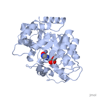
<table><tr><td colspan='2'>[[2ada]] is a 1 chain structure with sequence from [https://en.wikipedia.org/wiki/Mus_musculus Mus musculus]. This structure supersedes the now removed PDB entry [http://oca.weizmann.ac.il/oca-bin/send-pdb?obs=1&id=1ada 1ada]. Full crystallographic information is available from [http://oca.weizmann.ac.il/oca-bin/ocashort?id=2ADA OCA]. For a <b>guided tour on the structure components</b> use [https://proteopedia.org/fgij/fg.htm?mol=2ADA FirstGlance]. <br> | |||
</td></tr><tr id='method'><td class="sblockLbl"><b>[[Empirical_models|Method:]]</b></td><td class="sblockDat" id="methodDat">X-ray diffraction, [[Resolution|Resolution]] 2.4Å</td></tr> | |||
<tr id='ligand'><td class="sblockLbl"><b>[[Ligand|Ligands:]]</b></td><td class="sblockDat" id="ligandDat"><scene name='pdbligand=HPR:6-HYDROXY-7,8-DIHYDRO+PURINE+NUCLEOSIDE'>HPR</scene>, <scene name='pdbligand=ZN:ZINC+ION'>ZN</scene></td></tr> | |||
<tr id='resources'><td class="sblockLbl"><b>Resources:</b></td><td class="sblockDat"><span class='plainlinks'>[https://proteopedia.org/fgij/fg.htm?mol=2ada FirstGlance], [http://oca.weizmann.ac.il/oca-bin/ocaids?id=2ada OCA], [https://pdbe.org/2ada PDBe], [https://www.rcsb.org/pdb/explore.do?structureId=2ada RCSB], [https://www.ebi.ac.uk/pdbsum/2ada PDBsum], [https://prosat.h-its.org/prosat/prosatexe?pdbcode=2ada ProSAT]</span></td></tr> | |||
</table> | |||
== Function == | |||
[https://www.uniprot.org/uniprot/ADA_MOUSE ADA_MOUSE] Catalyzes the hydrolytic deamination of adenosine and 2-deoxyadenosine. Plays an important role in purine metabolism and in adenosine homeostasis. Modulates signaling by extracellular adenosine, and so contributes indirectly to cellular signaling events. Acts as a positive regulator of T-cell coactivation, by binding DPP4. Its interaction with DPP4 regulates lymphocyte-epithelial cell adhesion (By similarity). | |||
== Evolutionary Conservation == | |||
[[Image:Consurf_key_small.gif|200px|right]] | |||
Check<jmol> | |||
<jmolCheckbox> | |||
<scriptWhenChecked>; select protein; define ~consurf_to_do selected; consurf_initial_scene = true; script "/wiki/ConSurf/ad/2ada_consurf.spt"</scriptWhenChecked> | |||
<scriptWhenUnchecked>script /wiki/extensions/Proteopedia/spt/initialview01.spt</scriptWhenUnchecked> | |||
<text>to colour the structure by Evolutionary Conservation</text> | |||
</jmolCheckbox> | |||
</jmol>, as determined by [http://consurfdb.tau.ac.il/ ConSurfDB]. You may read the [[Conservation%2C_Evolutionary|explanation]] of the method and the full data available from [http://bental.tau.ac.il/new_ConSurfDB/main_output.php?pdb_ID=2ada ConSurf]. | |||
<div style="clear:both"></div> | |||
==See Also== | |||
*[[Adenosine deaminase 3D structures|Adenosine deaminase 3D structures]] | |||
__TOC__ | |||
== | </StructureSection> | ||
[[Category: Large Structures]] | |||
[[Category: | |||
[[Category: Mus musculus]] | [[Category: Mus musculus]] | ||
[[Category: Quiocho FA]] | |||
[[Category: Quiocho | [[Category: Wilson DK]] | ||
[[Category: Wilson | |||
Latest revision as of 12:12, 14 February 2024
ATOMIC STRUCTURE OF ADENOSINE DEAMINASE COMPLEXED WITH A TRANSITION-STATE ANALOG: UNDERSTANDING CATALYSIS AND IMMUNODEFICIENCY MUTATIONSATOMIC STRUCTURE OF ADENOSINE DEAMINASE COMPLEXED WITH A TRANSITION-STATE ANALOG: UNDERSTANDING CATALYSIS AND IMMUNODEFICIENCY MUTATIONS
Structural highlights
FunctionADA_MOUSE Catalyzes the hydrolytic deamination of adenosine and 2-deoxyadenosine. Plays an important role in purine metabolism and in adenosine homeostasis. Modulates signaling by extracellular adenosine, and so contributes indirectly to cellular signaling events. Acts as a positive regulator of T-cell coactivation, by binding DPP4. Its interaction with DPP4 regulates lymphocyte-epithelial cell adhesion (By similarity). Evolutionary Conservation Check, as determined by ConSurfDB. You may read the explanation of the method and the full data available from ConSurf. See Also |
| ||||||||||||||||||