1mbn: Difference between revisions
Jump to navigation
Jump to search
No edit summary |
No edit summary |
||
| (12 intermediate revisions by the same user not shown) | |||
| Line 1: | Line 1: | ||
< | ==The stereochemistry of the protein myoglobin== | ||
The | <StructureSection load='1mbn' size='340' side='right'caption='[[1mbn]], [[Resolution|resolution]] 2.00Å' scene=''> | ||
== Structural highlights == | |||
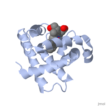
<table><tr><td colspan='2'>[[1mbn]] is a 1 chain structure with sequence from [https://en.wikipedia.org/wiki/Physeter_catodon Physeter catodon]. The January 2000 RCSB PDB [https://pdb.rcsb.org/pdb/static.do?p=education_discussion/molecule_of_the_month/index.html Molecule of the Month] feature on ''Myoglobin'' by David S. Goodsell is [https://dx.doi.org/10.2210/rcsb_pdb/mom_2000_1 10.2210/rcsb_pdb/mom_2000_1]. The October 2011 RCSB PDB [https://pdb.rcsb.org/pdb/static.do?p=education_discussion/molecule_of_the_month/index.html Molecule of the Month] feature on ''PDB Pioneers'' by David Goodsell is [https://dx.doi.org/10.2210/rcsb_pdb/mom_2011_10 10.2210/rcsb_pdb/mom_2011_10]. Full crystallographic information is available from [http://oca.weizmann.ac.il/oca-bin/ocashort?id=1MBN OCA]. For a <b>guided tour on the structure components</b> use [https://proteopedia.org/fgij/fg.htm?mol=1MBN FirstGlance]. <br> | |||
</td></tr><tr id='method'><td class="sblockLbl"><b>[[Empirical_models|Method:]]</b></td><td class="sblockDat" id="methodDat">X-ray diffraction, [[Resolution|Resolution]] 2Å</td></tr> | |||
<tr id='ligand'><td class="sblockLbl"><b>[[Ligand|Ligands:]]</b></td><td class="sblockDat" id="ligandDat"><scene name='pdbligand=HEM:PROTOPORPHYRIN+IX+CONTAINING+FE'>HEM</scene>, <scene name='pdbligand=OH:HYDROXIDE+ION'>OH</scene></td></tr> | |||
<tr id='resources'><td class="sblockLbl"><b>Resources:</b></td><td class="sblockDat"><span class='plainlinks'>[https://proteopedia.org/fgij/fg.htm?mol=1mbn FirstGlance], [http://oca.weizmann.ac.il/oca-bin/ocaids?id=1mbn OCA], [https://pdbe.org/1mbn PDBe], [https://www.rcsb.org/pdb/explore.do?structureId=1mbn RCSB], [https://www.ebi.ac.uk/pdbsum/1mbn PDBsum], [https://prosat.h-its.org/prosat/prosatexe?pdbcode=1mbn ProSAT]</span></td></tr> | |||
</table> | |||
== Function == | |||
[https://www.uniprot.org/uniprot/MYG_PHYMC MYG_PHYMC] Serves as a reserve supply of oxygen and facilitates the movement of oxygen within muscles. | |||
== Evolutionary Conservation == | |||
[[Image:Consurf_key_small.gif|200px|right]] | |||
Check<jmol> | |||
<jmolCheckbox> | |||
<scriptWhenChecked>; select protein; define ~consurf_to_do selected; consurf_initial_scene = true; script "/wiki/ConSurf/mb/1mbn_consurf.spt"</scriptWhenChecked> | |||
<scriptWhenUnchecked>script /wiki/extensions/Proteopedia/spt/initialview01.spt</scriptWhenUnchecked> | |||
<text>to colour the structure by Evolutionary Conservation</text> | |||
</jmolCheckbox> | |||
</jmol>, as determined by [http://consurfdb.tau.ac.il/ ConSurfDB]. You may read the [[Conservation%2C_Evolutionary|explanation]] of the method and the full data available from [http://bental.tau.ac.il/new_ConSurfDB/main_output.php?pdb_ID=1mbn ConSurf]. | |||
<div style="clear:both"></div> | |||
== | ==See Also== | ||
*[[Myoglobin 3D structures|Myoglobin 3D structures]] | |||
__TOC__ | |||
</StructureSection> | |||
[[Category: Large Structures]] | |||
[[Category: Myoglobin]] | [[Category: Myoglobin]] | ||
[[Category: PDB Pioneers]] | |||
[[Category: Physeter catodon]] | [[Category: Physeter catodon]] | ||
[[Category: | [[Category: RCSB PDB Molecule of the Month]] | ||
[[Category: | [[Category: Kendrew JC]] | ||
[[Category: | [[Category: Watson HC]] | ||
Latest revision as of 10:42, 14 February 2024
The stereochemistry of the protein myoglobinThe stereochemistry of the protein myoglobin
Structural highlights
FunctionMYG_PHYMC Serves as a reserve supply of oxygen and facilitates the movement of oxygen within muscles. Evolutionary Conservation Check, as determined by ConSurfDB. You may read the explanation of the method and the full data available from ConSurf. See Also |
| ||||||||||||||||||