1eyg: Difference between revisions
Jump to navigation
Jump to search
No edit summary |
No edit summary |
||
| (12 intermediate revisions by the same user not shown) | |||
| Line 1: | Line 1: | ||
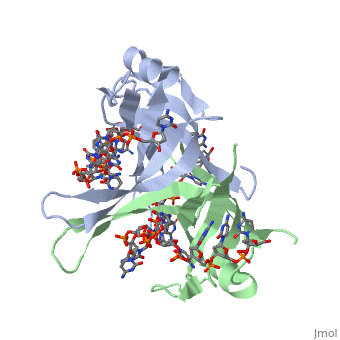
==Crystal structure of chymotryptic fragment of E. coli ssb bound to two 35-mer single strand DNAS== | |||
<StructureSection load='1eyg' size='340' side='right'caption='[[1eyg]], [[Resolution|resolution]] 2.80Å' scene=''> | |||
== Structural highlights == | |||
<table><tr><td colspan='2'>[[1eyg]] is a 6 chain structure with sequence from [https://en.wikipedia.org/wiki/Escherichia_coli Escherichia coli]. Full crystallographic information is available from [http://oca.weizmann.ac.il/oca-bin/ocashort?id=1EYG OCA]. For a <b>guided tour on the structure components</b> use [https://proteopedia.org/fgij/fg.htm?mol=1EYG FirstGlance]. <br> | |||
</td></tr><tr id='method'><td class="sblockLbl"><b>[[Empirical_models|Method:]]</b></td><td class="sblockDat" id="methodDat">X-ray diffraction, [[Resolution|Resolution]] 2.8Å</td></tr> | |||
<tr id='resources'><td class="sblockLbl"><b>Resources:</b></td><td class="sblockDat"><span class='plainlinks'>[https://proteopedia.org/fgij/fg.htm?mol=1eyg FirstGlance], [http://oca.weizmann.ac.il/oca-bin/ocaids?id=1eyg OCA], [https://pdbe.org/1eyg PDBe], [https://www.rcsb.org/pdb/explore.do?structureId=1eyg RCSB], [https://www.ebi.ac.uk/pdbsum/1eyg PDBsum], [https://prosat.h-its.org/prosat/prosatexe?pdbcode=1eyg ProSAT]</span></td></tr> | |||
| | </table> | ||
== Function == | |||
[https://www.uniprot.org/uniprot/SSB_ECOLI SSB_ECOLI] This protein is essential for replication of the chromosomes and its single-stranded DNA phages. It is also involved in DNA recombination and repair. | |||
== Evolutionary Conservation == | |||
[[Image:Consurf_key_small.gif|200px|right]] | |||
Check<jmol> | |||
<jmolCheckbox> | |||
<scriptWhenChecked>; select protein; define ~consurf_to_do selected; consurf_initial_scene = true; script "/wiki/ConSurf/ey/1eyg_consurf.spt"</scriptWhenChecked> | |||
<scriptWhenUnchecked>script /wiki/extensions/Proteopedia/spt/initialview01.spt</scriptWhenUnchecked> | |||
<text>to colour the structure by Evolutionary Conservation</text> | |||
</jmolCheckbox> | |||
</jmol>, as determined by [http://consurfdb.tau.ac.il/ ConSurfDB]. You may read the [[Conservation%2C_Evolutionary|explanation]] of the method and the full data available from [http://bental.tau.ac.il/new_ConSurfDB/main_output.php?pdb_ID=1eyg ConSurf]. | |||
<div style="clear:both"></div> | |||
==See Also== | |||
*[[Single-stranded DNA-binding protein 3D structures|Single-stranded DNA-binding protein 3D structures]] | |||
__TOC__ | |||
== | </StructureSection> | ||
[[Category: Escherichia coli]] | [[Category: Escherichia coli]] | ||
[[Category: | [[Category: Large Structures]] | ||
[[Category: Raghunathan | [[Category: Raghunathan S]] | ||
[[Category: Waksman | [[Category: Waksman G]] | ||
Latest revision as of 10:08, 7 February 2024
Crystal structure of chymotryptic fragment of E. coli ssb bound to two 35-mer single strand DNASCrystal structure of chymotryptic fragment of E. coli ssb bound to two 35-mer single strand DNAS
Structural highlights
FunctionSSB_ECOLI This protein is essential for replication of the chromosomes and its single-stranded DNA phages. It is also involved in DNA recombination and repair. Evolutionary Conservation Check, as determined by ConSurfDB. You may read the explanation of the method and the full data available from ConSurf. See Also |
| ||||||||||||||||