1atg: Difference between revisions
Jump to navigation
Jump to search
No edit summary |
No edit summary |
||
| (20 intermediate revisions by the same user not shown) | |||
| Line 1: | Line 1: | ||
== | ==AZOTOBACTER VINELANDII PERIPLASMIC MOLYBDATE-BINDING PROTEIN== | ||
<StructureSection load='1atg' size='340' side='right'caption='[[1atg]], [[Resolution|resolution]] 1.20Å' scene=''> | |||
== Structural highlights == | |||
<table><tr><td colspan='2'>[[1atg]] is a 1 chain structure with sequence from [https://en.wikipedia.org/wiki/Azotobacter_vinelandii Azotobacter vinelandii]. Full crystallographic information is available from [http://oca.weizmann.ac.il/oca-bin/ocashort?id=1ATG OCA]. For a <b>guided tour on the structure components</b> use [https://proteopedia.org/fgij/fg.htm?mol=1ATG FirstGlance]. <br> | |||
</td></tr><tr id='method'><td class="sblockLbl"><b>[[Empirical_models|Method:]]</b></td><td class="sblockDat" id="methodDat">X-ray diffraction, [[Resolution|Resolution]] 1.2Å</td></tr> | |||
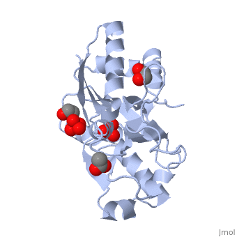
<tr id='ligand'><td class="sblockLbl"><b>[[Ligand|Ligands:]]</b></td><td class="sblockDat" id="ligandDat"><scene name='pdbligand=ACT:ACETATE+ION'>ACT</scene>, <scene name='pdbligand=EDO:1,2-ETHANEDIOL'>EDO</scene>, <scene name='pdbligand=SO4:SULFATE+ION'>SO4</scene>, <scene name='pdbligand=WO4:TUNGSTATE(VI)ION'>WO4</scene></td></tr> | |||
<tr id='resources'><td class="sblockLbl"><b>Resources:</b></td><td class="sblockDat"><span class='plainlinks'>[https://proteopedia.org/fgij/fg.htm?mol=1atg FirstGlance], [http://oca.weizmann.ac.il/oca-bin/ocaids?id=1atg OCA], [https://pdbe.org/1atg PDBe], [https://www.rcsb.org/pdb/explore.do?structureId=1atg RCSB], [https://www.ebi.ac.uk/pdbsum/1atg PDBsum], [https://prosat.h-its.org/prosat/prosatexe?pdbcode=1atg ProSAT]</span></td></tr> | |||
</table> | |||
== Function == | |||
[https://www.uniprot.org/uniprot/Q7SIH2_AZOVI Q7SIH2_AZOVI] | |||
== Evolutionary Conservation == | |||
[[Image:Consurf_key_small.gif|200px|right]] | |||
Check<jmol> | |||
<jmolCheckbox> | |||
<scriptWhenChecked>; select protein; define ~consurf_to_do selected; consurf_initial_scene = true; script "/wiki/ConSurf/at/1atg_consurf.spt"</scriptWhenChecked> | |||
<scriptWhenUnchecked>script /wiki/extensions/Proteopedia/spt/initialview01.spt</scriptWhenUnchecked> | |||
<text>to colour the structure by Evolutionary Conservation</text> | |||
</jmolCheckbox> | |||
</jmol>, as determined by [http://consurfdb.tau.ac.il/ ConSurfDB]. You may read the [[Conservation%2C_Evolutionary|explanation]] of the method and the full data available from [http://bental.tau.ac.il/new_ConSurfDB/main_output.php?pdb_ID=1atg ConSurf]. | |||
<div style="clear:both"></div> | |||
== | ==See Also== | ||
*[[ABC transporter 3D structures|ABC transporter 3D structures]] | |||
*[[ModG|ModG]] | |||
__TOC__ | |||
</StructureSection> | |||
[[Category: Azotobacter vinelandii]] | [[Category: Azotobacter vinelandii]] | ||
[[Category: | [[Category: Large Structures]] | ||
[[Category: Lawson | [[Category: Lawson DM]] | ||
[[Category: Mitchenall | [[Category: Mitchenall LA]] | ||
[[Category: Pau | [[Category: Pau RN]] | ||
[[Category: Williams | [[Category: Williams CEM]] | ||
Latest revision as of 09:33, 7 February 2024
AZOTOBACTER VINELANDII PERIPLASMIC MOLYBDATE-BINDING PROTEINAZOTOBACTER VINELANDII PERIPLASMIC MOLYBDATE-BINDING PROTEIN
Structural highlights
FunctionEvolutionary Conservation Check, as determined by ConSurfDB. You may read the explanation of the method and the full data available from ConSurf. See Also |
| ||||||||||||||||||