103l: Difference between revisions
Jump to navigation
Jump to search
No edit summary |
No edit summary |
||
| (19 intermediate revisions by the same user not shown) | |||
| Line 1: | Line 1: | ||
== | ==HOW AMINO-ACID INSERTIONS ARE ALLOWED IN AN ALPHA-HELIX OF T4 LYSOZYME== | ||
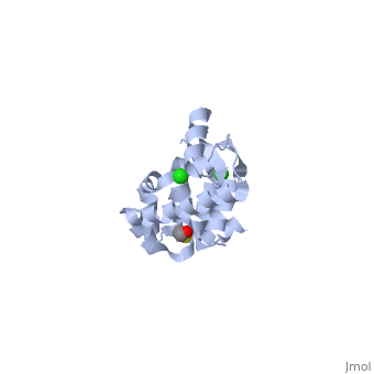
<StructureSection load='103l' size='340' side='right'caption='[[103l]], [[Resolution|resolution]] 1.90Å' scene=''> | |||
== Structural highlights == | |||
<table><tr><td colspan='2'>[[103l]] is a 1 chain structure with sequence from [https://en.wikipedia.org/wiki/Escherichia_virus_T4 Escherichia virus T4]. Full crystallographic information is available from [http://oca.weizmann.ac.il/oca-bin/ocashort?id=103L OCA]. For a <b>guided tour on the structure components</b> use [https://proteopedia.org/fgij/fg.htm?mol=103L FirstGlance]. <br> | |||
</td></tr><tr id='method'><td class="sblockLbl"><b>[[Empirical_models|Method:]]</b></td><td class="sblockDat" id="methodDat">X-ray diffraction, [[Resolution|Resolution]] 1.9Å</td></tr> | |||
<tr id='ligand'><td class="sblockLbl"><b>[[Ligand|Ligands:]]</b></td><td class="sblockDat" id="ligandDat"><scene name='pdbligand=BME:BETA-MERCAPTOETHANOL'>BME</scene>, <scene name='pdbligand=CL:CHLORIDE+ION'>CL</scene></td></tr> | |||
<tr id='resources'><td class="sblockLbl"><b>Resources:</b></td><td class="sblockDat"><span class='plainlinks'>[https://proteopedia.org/fgij/fg.htm?mol=103l FirstGlance], [http://oca.weizmann.ac.il/oca-bin/ocaids?id=103l OCA], [https://pdbe.org/103l PDBe], [https://www.rcsb.org/pdb/explore.do?structureId=103l RCSB], [https://www.ebi.ac.uk/pdbsum/103l PDBsum], [https://prosat.h-its.org/prosat/prosatexe?pdbcode=103l ProSAT]</span></td></tr> | |||
</table> | |||
== Function == | |||
[https://www.uniprot.org/uniprot/ENLYS_BPT4 ENLYS_BPT4] Endolysin with lysozyme activity that degrades host peptidoglycans and participates with the holin and spanin proteins in the sequential events which lead to the programmed host cell lysis releasing the mature viral particles. Once the holin has permeabilized the host cell membrane, the endolysin can reach the periplasm and break down the peptidoglycan layer.<ref>PMID:22389108</ref> | |||
== Evolutionary Conservation == | |||
[[Image:Consurf_key_small.gif|200px|right]] | |||
Check<jmol> | |||
<jmolCheckbox> | |||
<scriptWhenChecked>; select protein; define ~consurf_to_do selected; consurf_initial_scene = true; script "/wiki/ConSurf/03/103l_consurf.spt"</scriptWhenChecked> | |||
<scriptWhenUnchecked>script /wiki/extensions/Proteopedia/spt/initialview01.spt</scriptWhenUnchecked> | |||
<text>to colour the structure by Evolutionary Conservation</text> | |||
</jmolCheckbox> | |||
</jmol>, as determined by [http://consurfdb.tau.ac.il/ ConSurfDB]. You may read the [[Conservation%2C_Evolutionary|explanation]] of the method and the full data available from [http://bental.tau.ac.il/new_ConSurfDB/main_output.php?pdb_ID=103l ConSurf]. | |||
<div style="clear:both"></div> | |||
== | ==See Also== | ||
*[[Lysozyme 3D structures|Lysozyme 3D structures]] | |||
== References == | |||
== | <references/> | ||
__TOC__ | |||
[[Category: | </StructureSection> | ||
[[Category: | [[Category: Escherichia virus T4]] | ||
[[Category: Heinz | [[Category: Large Structures]] | ||
[[Category: Matthews | [[Category: Heinz DW]] | ||
[[Category: Matthews BW]] | |||
Latest revision as of 09:23, 7 February 2024
HOW AMINO-ACID INSERTIONS ARE ALLOWED IN AN ALPHA-HELIX OF T4 LYSOZYMEHOW AMINO-ACID INSERTIONS ARE ALLOWED IN AN ALPHA-HELIX OF T4 LYSOZYME
Structural highlights
FunctionENLYS_BPT4 Endolysin with lysozyme activity that degrades host peptidoglycans and participates with the holin and spanin proteins in the sequential events which lead to the programmed host cell lysis releasing the mature viral particles. Once the holin has permeabilized the host cell membrane, the endolysin can reach the periplasm and break down the peptidoglycan layer.[1] Evolutionary Conservation Check, as determined by ConSurfDB. You may read the explanation of the method and the full data available from ConSurf. See AlsoReferences
|
| ||||||||||||||||||