2cfp: Difference between revisions
No edit summary |
No edit summary |
||
| (10 intermediate revisions by the same user not shown) | |||
| Line 1: | Line 1: | ||
< | ==Sugar Free Lactose Permease at acidic pH== | ||
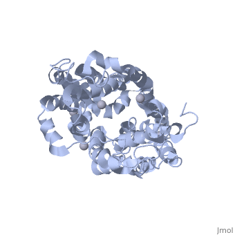
<StructureSection load='2cfp' size='340' side='right'caption='[[2cfp]], [[Resolution|resolution]] 3.30Å' scene=''> | |||
You may | == Structural highlights == | ||
<table><tr><td colspan='2'>[[2cfp]] is a 1 chain structure with sequence from [https://en.wikipedia.org/wiki/Escherichia_coli Escherichia coli]. Full crystallographic information is available from [http://oca.weizmann.ac.il/oca-bin/ocashort?id=2CFP OCA]. For a <b>guided tour on the structure components</b> use [https://proteopedia.org/fgij/fg.htm?mol=2CFP FirstGlance]. <br> | |||
</td></tr><tr id='method'><td class="sblockLbl"><b>[[Empirical_models|Method:]]</b></td><td class="sblockDat" id="methodDat">X-ray diffraction, [[Resolution|Resolution]] 3.3Å</td></tr> | |||
<tr id='ligand'><td class="sblockLbl"><b>[[Ligand|Ligands:]]</b></td><td class="sblockDat" id="ligandDat"><scene name='pdbligand=HG:MERCURY+(II)+ION'>HG</scene></td></tr> | |||
<tr id='resources'><td class="sblockLbl"><b>Resources:</b></td><td class="sblockDat"><span class='plainlinks'>[https://proteopedia.org/fgij/fg.htm?mol=2cfp FirstGlance], [http://oca.weizmann.ac.il/oca-bin/ocaids?id=2cfp OCA], [https://pdbe.org/2cfp PDBe], [https://www.rcsb.org/pdb/explore.do?structureId=2cfp RCSB], [https://www.ebi.ac.uk/pdbsum/2cfp PDBsum], [https://prosat.h-its.org/prosat/prosatexe?pdbcode=2cfp ProSAT]</span></td></tr> | |||
</table> | |||
== Function == | |||
[https://www.uniprot.org/uniprot/LACY_ECOLI LACY_ECOLI] Responsible for transport of beta-galactosides into the cell, with the concomitant import of a proton (symport system). | |||
== Evolutionary Conservation == | |||
[[Image:Consurf_key_small.gif|200px|right]] | |||
Check<jmol> | |||
<jmolCheckbox> | |||
<scriptWhenChecked>; select protein; define ~consurf_to_do selected; consurf_initial_scene = true; script "/wiki/ConSurf/cf/2cfp_consurf.spt"</scriptWhenChecked> | |||
<scriptWhenUnchecked>script /wiki/extensions/Proteopedia/spt/initialview01.spt</scriptWhenUnchecked> | |||
<text>to colour the structure by Evolutionary Conservation</text> | |||
</jmolCheckbox> | |||
</jmol>, as determined by [http://consurfdb.tau.ac.il/ ConSurfDB]. You may read the [[Conservation%2C_Evolutionary|explanation]] of the method and the full data available from [http://bental.tau.ac.il/new_ConSurfDB/main_output.php?pdb_ID=2cfp ConSurf]. | |||
<div style="clear:both"></div> | |||
<div style="background-color:#fffaf0;"> | |||
== Publication Abstract from PubMed == | |||
Cation-coupled active transport is an essential cellular process found ubiquitously in all living organisms. Here, we present two novel ligand-free X-ray structures of the lactose permease (LacY) of Escherichia coli determined at acidic and neutral pH, and propose a model for the mechanism of coupling between lactose and H+ translocation. No sugar-binding site is observed in the absence of ligand, and deprotonation of the key residue Glu269 is associated with ligand binding. Thus, substrate induces formation of the sugar-binding site, as well as the initial step in H+ transduction. | |||
Structural evidence for induced fit and a mechanism for sugar/H+ symport in LacY.,Mirza O, Guan L, Verner G, Iwata S, Kaback HR EMBO J. 2006 Mar 22;25(6):1177-83. Epub 2006 Mar 9. PMID:16525509<ref>PMID:16525509</ref> | |||
From MEDLINE®/PubMed®, a database of the U.S. National Library of Medicine.<br> | |||
</div> | |||
<div class="pdbe-citations 2cfp" style="background-color:#fffaf0;"></div> | |||
==See Also== | |||
*[[Lactose Permease|Lactose Permease]] | |||
== References == | |||
<references/> | |||
__TOC__ | |||
</StructureSection> | |||
== | |||
== | |||
< | |||
[[Category: Escherichia coli]] | [[Category: Escherichia coli]] | ||
[[Category: Guan | [[Category: Large Structures]] | ||
[[Category: Iwata | [[Category: Guan L]] | ||
[[Category: Kaback | [[Category: Iwata S]] | ||
[[Category: Mirza | [[Category: Kaback HR]] | ||
[[Category: Verner | [[Category: Mirza O]] | ||
[[Category: Verner G]] | |||
Latest revision as of 17:16, 13 December 2023
Sugar Free Lactose Permease at acidic pHSugar Free Lactose Permease at acidic pH
Structural highlights
FunctionLACY_ECOLI Responsible for transport of beta-galactosides into the cell, with the concomitant import of a proton (symport system). Evolutionary Conservation Check, as determined by ConSurfDB. You may read the explanation of the method and the full data available from ConSurf. Publication Abstract from PubMedCation-coupled active transport is an essential cellular process found ubiquitously in all living organisms. Here, we present two novel ligand-free X-ray structures of the lactose permease (LacY) of Escherichia coli determined at acidic and neutral pH, and propose a model for the mechanism of coupling between lactose and H+ translocation. No sugar-binding site is observed in the absence of ligand, and deprotonation of the key residue Glu269 is associated with ligand binding. Thus, substrate induces formation of the sugar-binding site, as well as the initial step in H+ transduction. Structural evidence for induced fit and a mechanism for sugar/H+ symport in LacY.,Mirza O, Guan L, Verner G, Iwata S, Kaback HR EMBO J. 2006 Mar 22;25(6):1177-83. Epub 2006 Mar 9. PMID:16525509[1] From MEDLINE®/PubMed®, a database of the U.S. National Library of Medicine. See AlsoReferences |
| ||||||||||||||||||