Nuclear receptor: Difference between revisions
Jump to navigation
Jump to search
No edit summary |
No edit summary |
||
| (8 intermediate revisions by the same user not shown) | |||
| Line 5: | Line 5: | ||
</ref> | </ref> | ||
SEE [[Nuclear receptors]] | |||
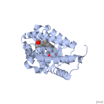
<StructureSection load='1ie8' size='350' side='right' caption='Structure of the LBD of a VDR (vitamin D receptor) complexed to 1alpha,25(OH)(2)D(3) and the 20-epi analogs(PDB entry [[1ie8]])' scene=''> | <StructureSection load='1ie8' size='350' side='right' caption='Structure of the LBD of a VDR (vitamin D receptor) complexed to 1alpha,25(OH)(2)D(3) and the 20-epi analogs(PDB entry [[1ie8]])' scene=''> | ||
| Line 18: | Line 15: | ||
arranged in a three layered sandwich, and a <scene name='Nuclear_receptor/Nr-lbd-beta/1'>beta-sheet</scene>. | arranged in a three layered sandwich, and a <scene name='Nuclear_receptor/Nr-lbd-beta/1'>beta-sheet</scene>. | ||
</StructureSection> | </StructureSection> | ||
<references/> | <references/> | ||
Latest revision as of 19:36, 25 January 2021
the nuclear receptors facts book [1]
The structure of the LBD is strongly conserved throughout the familly of nuclear receptors. It is a globular domain, mainly alpha composed of arranged in a three layered sandwich, and a .
|
| ||||||||||
- ↑ Vincent Laudet, Hinrich Gronemeyer, "The nuclear receptor factsbook", Publisher Academic Press, 2002